43 liver histology diagram
When the process involves the liver diffusely and is accompanied by regenerative nodule formation, it is called cirrhosis ("end‐stage liver"). A trichrome stain is useful in confirming the presence of fibrosis. Examples of a few grades and stages of CH are illustrated in Figs. Figs.1, 1, ,2, 2, ,3, 3, and and4 4. Blood supply and innervation of the liver Author: Alexandra Sieroslawska MD • Reviewer: Dimitrios Mytilinaios MD, PhD Last reviewed: October 28, 2021 Reading time: 10 minutes The liver is the largest visceral tissue mass in the human body and is located in the upper right quadrant of the abdomen.It is a multifunctional accessory to the gastrointestinal tract and performs such duties as ...
Terminology. An accessory hepatic artery is one which arises from an anomalous origin and supplies a portion of the liver along with another artery.. A replaced hepatic artery is one which arises from an anomalous origin and supplies a portion of the liver solely.. Variant anatomy. In general, the common hepatic artery may arise from the abdominal aorta or superior mesenteric artery (SMA) and ...

Liver histology diagram
Veterinary students and farm owners might know the external and internal goat anatomy to maintain good health and increase the productivity of a herd.I will discuss the external and internal anatomical facts of a goat here in this article. This brief outline of the goat's anatomy is a starting point for those who want to keep the goat. Liver histology Author: Adrian Rad BSc (Hons) • Reviewer: Uruj Zehra MBBS, MPhil, PhD Last reviewed: October 04, 2021 Reading time: 14 minutes The liver is the largest internal organ of the human body, weighing approximately 1.5 kg. Embryologically it develops from the foregut and it spans the upper right and part of left abdominal quadrants. . Anatomically the liver consists of four lobes ... Importantly, restoration of SLU7 expression protected against chronic liver damage. AAV-SLU7 significantly reduced serum transaminases, improved liver histology, inhibited hepatic collagen expression and accumulation, and suppressed HSC activation (Fig. 7A-C), altogether translating into a reduced loss of body weight (Supporting Fig. S6C).
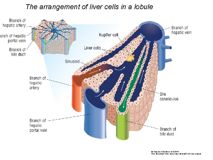
Liver histology diagram. The liver is a critical organ in the human body that is responsible for an array of functions that help support metabolism, immunity, digestion, detoxification, vitamin storage among other functions. It comprises around 2% of an adult's body weight. The liver is a unique organ due to its dual blood supply from the portal vein (approximately 75%) and the hepatic artery (approximately 25%). Full labeled anatomical diagrams - Anatomy of the abdomen and digestive system: these general diagrams show the digestive system, with the major human anatomical structures labeled (mouth, tongue, oral cavity, teeth, buccal glands, throat, pharynx, oesophagus, stomach, small intestine, large intestine, liver, gall bladder and pancreas). #2. Best guide to learn liver histology in details. Do you want to learn the details anatomy of lung from different animals? You may find details anatomy of lung here in this blog. Conclusion. Hope you got a better idea about lung histology. Please watch the video carefully and learn from lung histology diagram. In pancreas histology of animal, you will find the both endocrine and exocrine parts. The exocrine part forms the major portion of pancreas and consists of closely packed serous acini with zymogenic cells. Again, the endocrine part consists of pancreatic islets of Langerhans which is located within the masses of serous acini.
You might also read other different article related to histology of respiratory organs from anatomy learner - #1. Histological features of animal lung with slide image and labeled diagram #2. Identification of epiglottis under light microscope. Conclusion. This is the best guide to learn trachea histology with slide images and labeled diagram. An organoid model for ARPKD liver pathology. PKHD1 mutations that cause amino acid substitutions are generally associated with a non-lethal presentation, while neonatal death tends to be ... See more ideas about liver, histology slides, liver anatomy. ... liver diagram lobule - Google Search What Is Living, Bile Duct, Liver Cancer,. Normal spleen histology (diagram). Like every other organ, the spleen consists of stroma and parenchyma. The stroma of the spleen is composed mainly of a network of reticular connective tissue.This mesh provides support for blood cells and cells of the immune system (lymphocytes, macrophages, and dendritic cells). The parenchyma of the spleen is divided into two functionally and ...
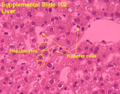
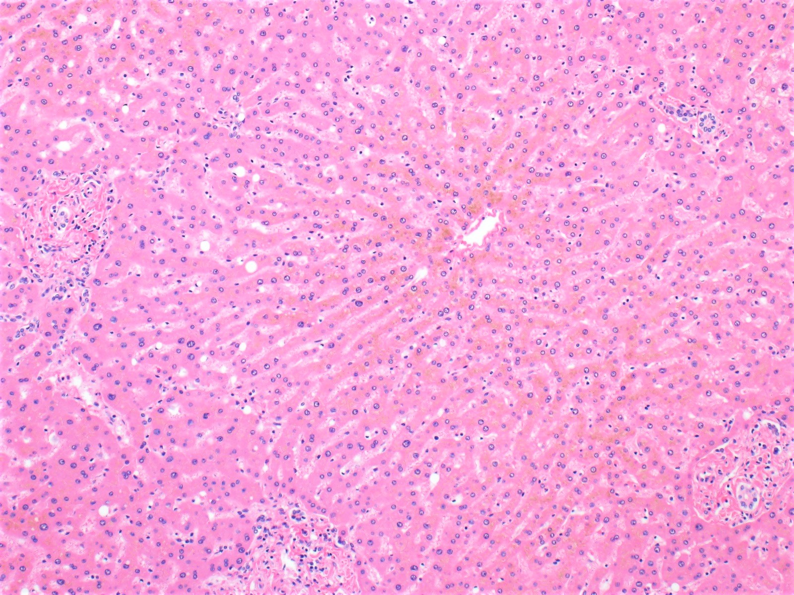
Liver The liver is the largest gland and chief metabolic organ of the body. It can be regarded as a huge biosynthetic chemical factory that synthesizes large complex molecules from the material transported to this organ by the blood. The chief functions of the liver can be grouped into five main categories as given below: […] By histology, these cells have foamy cytoplasm, which results from the surfactant that is stored as lamellar bodies. Type II pneumocytes are also mitotically active and can replace the easily damaged type I pneumocytes. Type II pneumocyte cells can be recognized by their rounded shapes that bulge into the alveolar space. The Human Digestive System Definition. The human digestive system is the collective name used to describe the alimentary canal, some accessory organs, and a variety of digestive processes that take place at different levels in the canal to prepare food eaten in the diet for absorption. See more ideas about liver, histology slides, bile duct. Child's Digestive System from childrensgimd.com Liver diagram illustrations & vectors. Where is the liver located in the human body diagram liver picture diagram locating liver pain the body uses pain as its means of. See more ideas about liver, histology slides, bile duct.

Liver Organization
Human liver wedge biopsies measuring ~ 1 cm × 1 cm × 1 cm were fixed with 1.5% glutaraldehyde in 0.067 M sodium cacodylate buffer (pH 7.4) (primary fixative) by means of injection perfusion fixation as previously described in detail 47. Following the injection of the fixative which induces discolouration and hardening of the soft tissue ...
Histology At Siu Liver
The liver panel is a test with multiple measurements that help to assess the health and function of the liver. The test is conducted with a blood sample that is normally taken from a vein in your arm. A liver panel can be used to help diagnose and monitor liver diseases.
Liver Histology Prac Looking At Liver Slides Starter
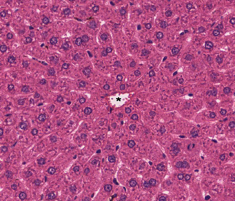
Start studying liver histology 100x (Lobule). Learn vocabulary, terms, and more with flashcards, games, and other study tools.

Association Between Magnetic Resonance Imaging Proton Density Fat Fraction And Liver Histology Features In Patients With Nonalcoholic Fatty Liver Disease Or Nonalcoholic Steatohepatitis Gastroenterology
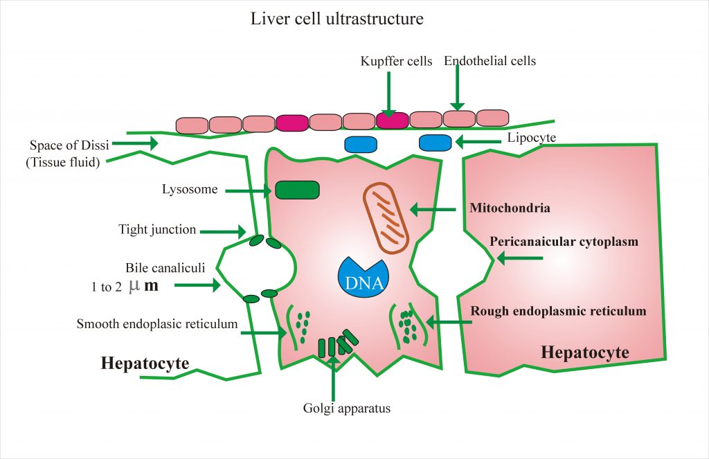
Feb 19, 2020 — These may play an active role in portal hypertension. This ultrastructure of the liver cell can be elaborated better by the following diagram.

Nutmeg Liver Histopathology Guru
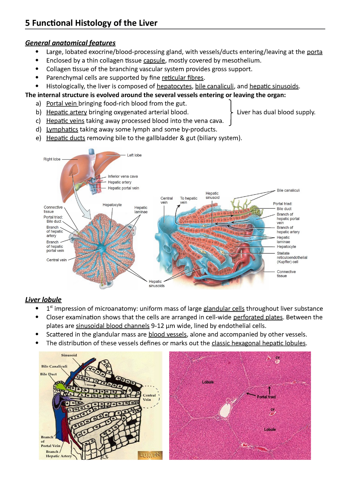
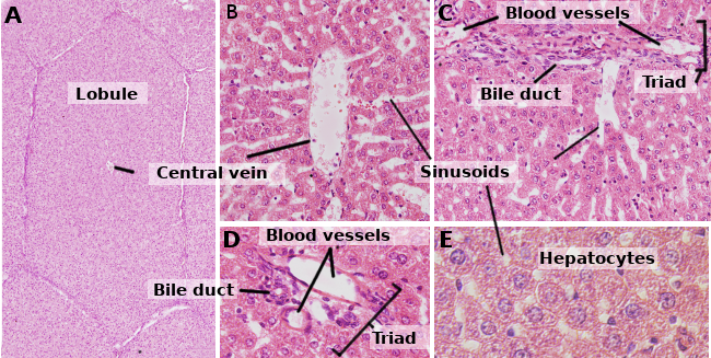
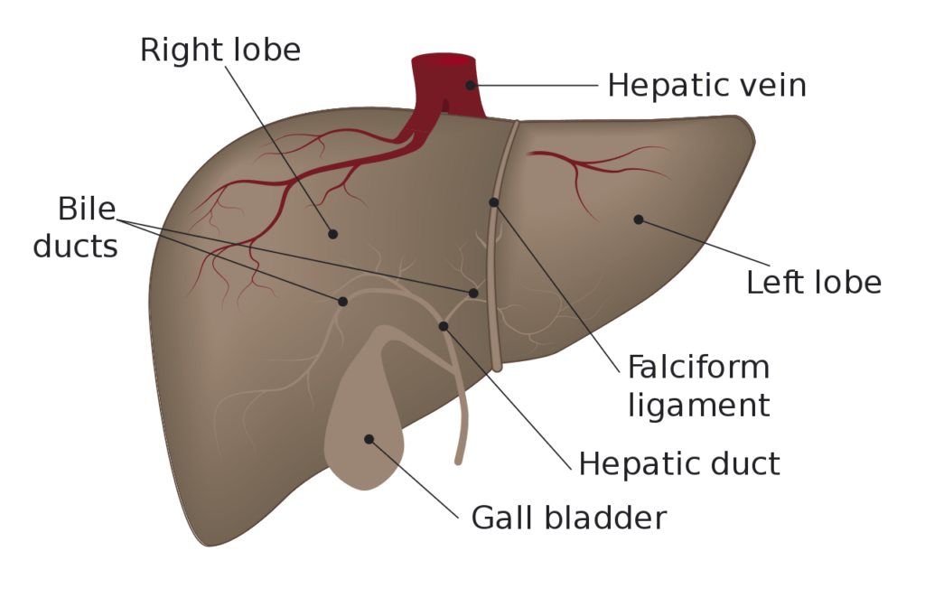
Gross anatomy. The liver is an irregular, wedge-shaped organ that lies below the diaphragm in the right upper quadrant of the abdominal cavity and is in close approximation with the diaphragm, stomach and the gallbladder.It is largely covered by the costal cartilages.. The liver is made of several functional units called lobules, which in turn can be subdivided into smaller units called sinusoids.

Liver Histology Diagram Diagram Quizlet
Liver and intrahepatic bile ducts - nontumor - Normal histology. The liver is the largest solid organ and is located in the right upper quadrant of the abdomen.
5 Anatomy And Functional Histology Of The Liver 5 Functional Histology Of The Liver General Studocu
Reticular tissue is a special type of connective tissue that predominates in various locations that have a high cellular content. It has a branched and mesh-like pattern, often called reticulum, due to the arrangement of reticular fibers (reticulin).These fibers are actually type III collagen fibrils.In comparison to the predominant type I collagen, type III fibrils are narrower, do not form ...

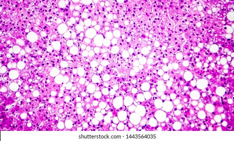
Histopathology Drawings Fatty Liver Facebook
The liver has a thin capsule of dense connective tissue, and a visceral (inferior) layer of peritoneal mesothelium, and is divided into left and right lobes.
Liver Gallbladder And Pancreas Histology
Liver, pancreas, and gallbladder microscope slides. From the liver histology slide, you might identify the polygonal hepatic lobules, hepatic cords, portal tried central vein and other different vital structures. You will find the detailed guide on liver histology here in anatomy learner.
96291123 Style Medical 3 Histology 1 Piece Powerpoint Presentation Diagram Infographic Slide Powerpoint Presentation Designs Slide Ppt Graphics Presentation Template Designs
Liver pancreas stomach diagram. An important organ of the digestive system the liver is located below the diaphragm and to the right of the stomach. In humans it is located in the right upper quadrant of the abdomen below the diaphragmIts other roles in metabolism include the regulation of glycogen storage decomposition of red blood cells and.

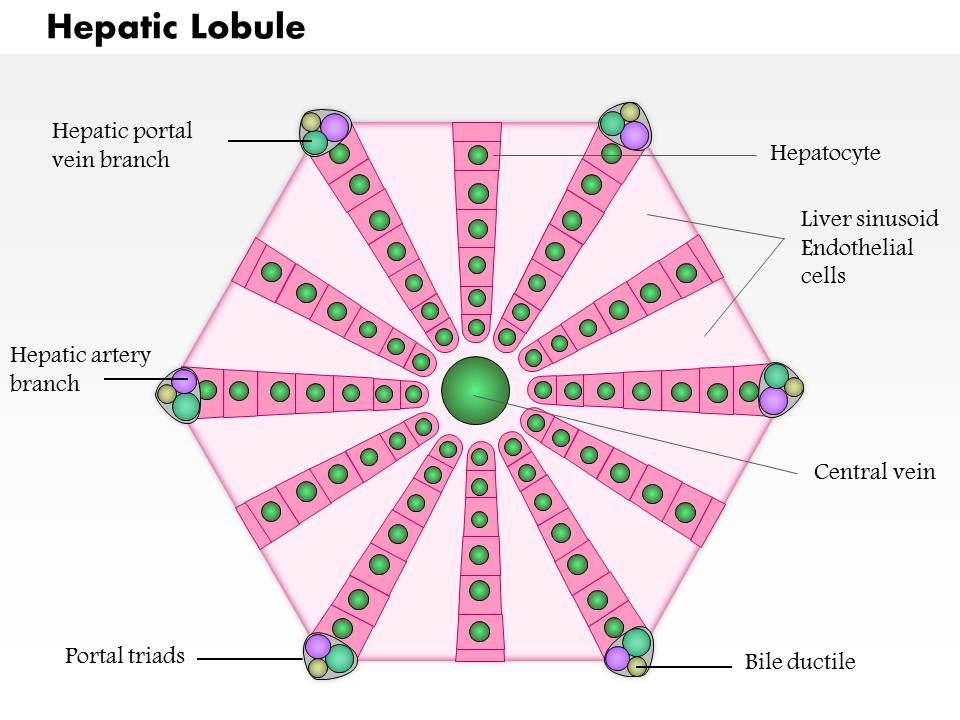
Histology Slides Database Histological Diagram Of Hepatic Lobule
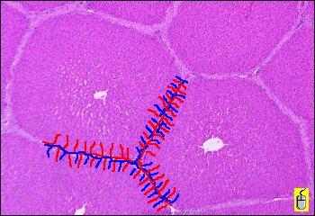
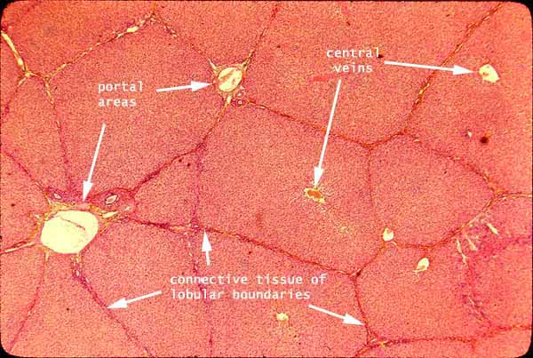
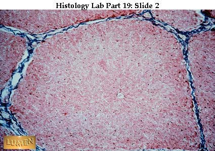
Histology of liver: Each lobe of liver consists of a large number of hepatic lobules that are separated from each other by a thin layer of connective tissue called septa or Glisson's capsule . In the liver of rabbit and human beings, septa demarcating the lobules are incomplete and not clearly marked off.

Histology Of The Liver
by GM Petcoff · 2006 · Cited by 47 — The liver is a mixed gland surrounded by a thin capsule of connective tissue, the Glisson capsule, dividing the parenchyma into lobules and lobuli. The ...
Liver Histology Labpedia Net
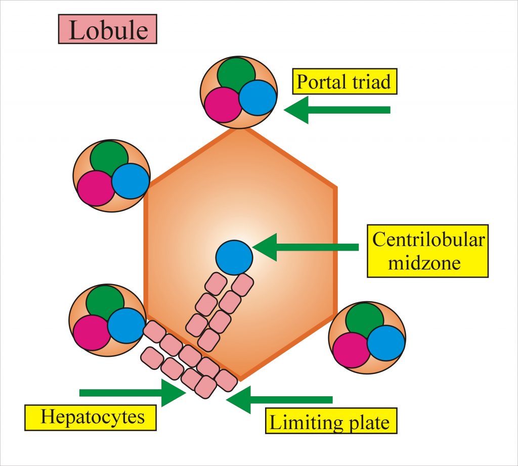
10. Classical Hepatic Lobule • Each lobule consists of a hexagonal mass of liver cells. ... Central axis occupied by: Central vein. ... Is an independent venous ...

Histology Of Liver Quiz
In the case of the liver, the roads are connective tissue septae which convey vascular and biliary traffic, and the clusters of houses are cord-like ...
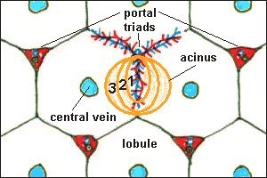
Hepatic Histology The Acinus
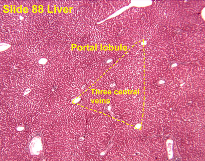
The liver acinus defines the liver based on its metabolic function and is center around its nutrient supply (distributing veins). It is the shape of a diamond with two central veins at 2 points ...
Arppress Org
"Centroid" approach for obtaining a Voronoi diagram based on the location of central veins in a section of normal porcine liver. The process starts with the centrilobular/zone 3 marker ...

Liver Histology Ppt Download
The liver is a large essential organ found in the upper right quadrant of the abdomen.It is a multifunctional accessory to the gastrointestinal tract and performs such duties as detoxification, protein synthesis, biochemical production and nutrient storage to name but a few. It is the largest gland in the human body, weighing in at approximately 1.5 kilograms.

Medical Liver Biopsy Background Indications Procedure And Histopathology Frontline Gastroenterology
Importantly, restoration of SLU7 expression protected against chronic liver damage. AAV-SLU7 significantly reduced serum transaminases, improved liver histology, inhibited hepatic collagen expression and accumulation, and suppressed HSC activation (Fig. 7A-C), altogether translating into a reduced loss of body weight (Supporting Fig. S6C).
Liver Histology Structure Cells And Characteristics Kenhub
Liver histology Author: Adrian Rad BSc (Hons) • Reviewer: Uruj Zehra MBBS, MPhil, PhD Last reviewed: October 04, 2021 Reading time: 14 minutes The liver is the largest internal organ of the human body, weighing approximately 1.5 kg. Embryologically it develops from the foregut and it spans the upper right and part of left abdominal quadrants. . Anatomically the liver consists of four lobes ...

Liver Histology Diagram Quizlet
Veterinary students and farm owners might know the external and internal goat anatomy to maintain good health and increase the productivity of a herd.I will discuss the external and internal anatomical facts of a goat here in this article. This brief outline of the goat's anatomy is a starting point for those who want to keep the goat.

Liver Histology Sciencekeys

Molecular And Histological Correlations In Liver Cancer Journal Of Hepatology
Liver Histology Images Stock Photos Vectors Shutterstock
Hepatic Histology The Acinus
Liver Gall Bladder Pancreas
Cell Types Hepatocyte Atlas Of Plant And Animal Histology
Liver Histology Labpedia Net
Pathology Outlines Histology
Liver Anatomy Histology And Functions Online Science Notes

Liver Histology Diagram Gilson Biology Knowledge Facebook
Histology At Siu

Group 1 Normal Histology Of Mouse Liver At 20x Magnification Download Scientific Diagram
Labeled

Histology Digestion Lab Pig Liver
Histology Slides 1
Liver Gall Bladder Pancreas
1

89 Histology Liver Ideas Liver Histology Slides Liver Anatomy

Histology Of The Liver Youtube

The Liver Medical Student Education Tissupath

Hepatic Pathology

10 Liver Ideas طب مدرسة علم
A Schematic Of A Typical Liver Lobule Adapted Under Cc By Nc 4 0 From Download Scientific Diagram


Komentar
Posting Komentar