41 cervical plexus diagram
Science Anatomy and Physiology Q&A Library 1. Damage to the cervical nerve plexus can cause problems with A) Breathing B) Clawhand C) Footdrop D) Wristdrop 2. Each spinal nerve divides into a dorsal and ventral A) Ganglion B) Plexus c) Ramus D) Tracts 3. Variability in brachial plexus anatomy can also possibly explain some of the variation in the clinical presentation of radicular symptoms. The brachial plexus is comprised of the ventral rami of the C5-T1 spinal roots. Based on anatomic dissections of the plexus, a "typical" plexus is only observed 37-77% of the time.
From the case: Cervical plexus (diagram) Diagram. Nerve to rectus capitis lateralis, longus capitis and rectus capitis anterior. Nerve to scalenes and levator scapulae. From the case: Cervical plexus (diagram) Diagram. Nerve to scalenes and levator scapulae. Phrenic nerve. From the case: Cervical plexus (diagram)
Cervical plexus diagram
Nerves of the Cervical Plexus - Labeling Diagram. Nerves of the Lower Limb - Labeling Diagram. Nerves of the Lumbar Plexus - Labeling Diagram. Nerves of the Sacral Plexus - Labeling Diagram. Nerves of the Upper Limb - Labeling Diagram. Nervous System - Label the Parts. ADVERTISEMENTS: In this article we will discuss about the dissection of rat. Also learn about:- 1. Dissection of Alimentary System 2. Dissection of Circulatory System 3. Dissection of Venous System 4. The Arterial System 5. Dissection of Cranial Nerves 6. Dissection of Brain 7. Dissection of Neck Region 8. Dissection of Urinogenital System 9. The […] cervical plexus. I am formed by the ventral rami of the first four cervical nerves who am I. again you are the cervical plexus. most branches are_____nerves that supply only the skin. cutaneous. I transmit sensory impulses from the skin of the neck the ear area the back of the head and the shoulder who am I.
Cervical plexus diagram. By convention, the cervical vertebrae are numbered, with the first one (C1) closest to the skull and higher numbered vertebrae (C2–C7) proceeding away from the skull and down the spine. The general characteristics of the third through sixth cervical vertebrae are described here. The first, second, and seventh vertebrae are extraordinary, and are detailed later. Sacral Plexus Diagram. Here are a number of highest rated Sacral Plexus Diagram pictures upon internet. We identified it from well-behaved source. Its submitted by government in the best field. We acknowledge this kind of Sacral Plexus Diagram graphic could possibly be the most trending subject similar to we allowance it in google lead or facebook. The phrenic nerve is a mixed motor/sensory nerve which originates from the C3-C5 spinal nerves in the neck. The nerve is important for breathing because it provides exclusive motor control of the diaphragm, the primary muscle of respiration. In humans, the right and left phrenic nerves are primarily supplied by the C4 spinal nerve, but there is also contribution from the C3 and C5 spinal nerves. The nerves of the cervical plexus supply the back of the head, the neck and the shoulders. Brachial Plexus The arrangement of nerve fibers formed by the ventral rami of the lower cervical and upper thoracic nerve root, precisely between the nerve roots of the 5th cervical and 1st thoracic vertebra, is known as the brachial plexus. It runs ...
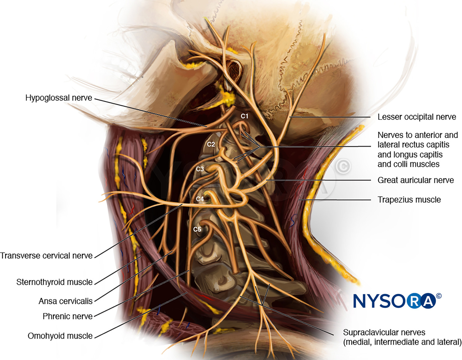
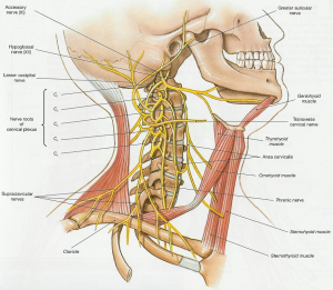
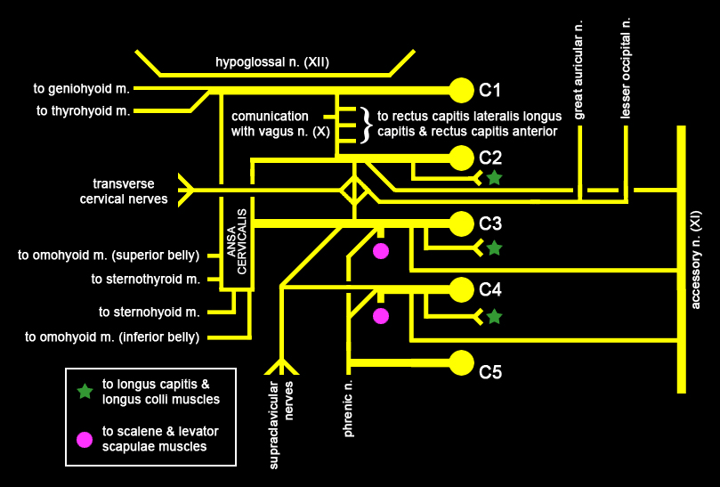
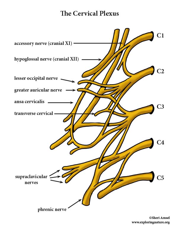
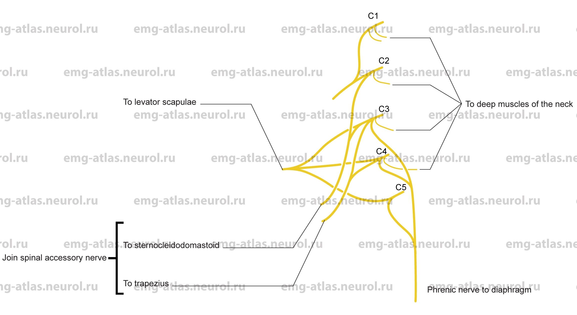
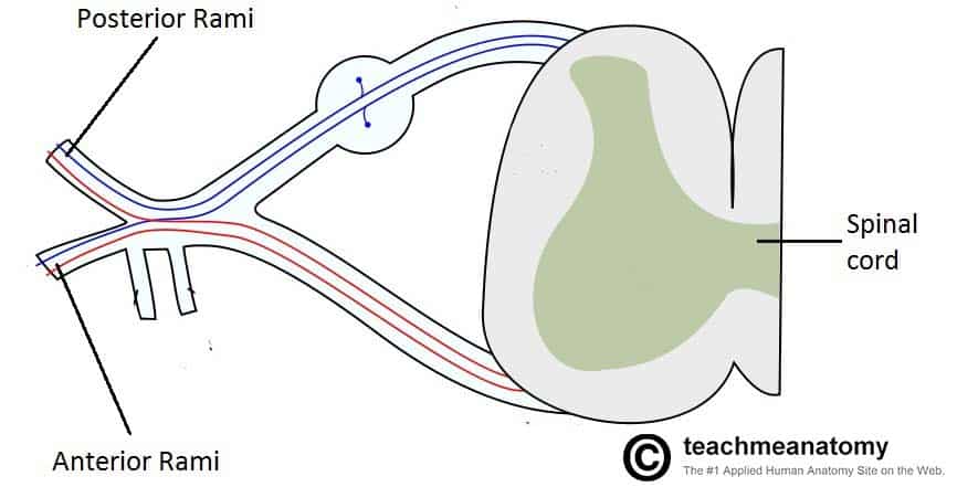
The cervical nerves consist of eight paired nerves that are a part of the peripheral nervous system. They emerge from the spinal cord through the seven cervical vertebrae. ... The brachial plexus ... Cervical Plexus This plexus is located underneath the sternocleidomastoid muscle (from C1 - C4). Most of the branches innervate the skin of neck and deep neck muscles. The Phrenic nerve (C3, C4, C5) gets special mention that innervates the top of the diaphragm (after traveling down through thoracic cavity, along either side of the heart). Cervical Plexus (Complete Diagram) © 2001, Allan Forsman The cervical plexus can be described as a web of nerves. A plexus is a combination of nerves. The cervical plexus is formed by the merging of the anterior portion of spinal nerves C1 through C4 and part of C5. There is some confusing terminology when it comes to anterior and posterior sections of spinal nerves. All spinal nerves are composed of the merging of anterior (ventral, front, motor) and posterior (dorsal, back, sensory) nerve rootsthat emerge from the spine. Once the spinal nerves form from their anterior and posterior components, each spinal nerve then divides again into an anterior and a posterior branch (rami). Anterior and posterior rami of any specific spinal nerve do not necessarily follow the same path. Anterior and posterior rami can be motor nerves, sensory nerves, or both. The cervical plexus arises from the anterior rami of the corresponding cervical spinal nerves.
The cervical plexus is a complex neurologic structure located within the head and neck. The large portion of the cervical plexus is the communication between the anterior divisions of C1 through C4 nerves. While the plexus itself can be complex, it is essential for practitioners to understand the significant motor and sensory functions of the cervical plexus as this information can provide ... Cervical spinal nerves Thoracic T spinal nerves Lumbar spinal nerves Sacral spinal nerves 2 Coccygeal Filum terminale nerve (Co 1) (in coccygeal ligament) Cauda equina Inferior tip of spinal cord Conus medullaris Lumbosacral enlargement Posterior median sulcus Cervical enlargement C 1 C 2 C 3 C 4 C 5 C 6 C 7 C 8 T 1 T 2 T 3 T 4 T 5 T 6 T 7 8 T ... Correction: The Lesser Occipital nerve arises from C2 and C3, not C2 only. Draw it in between C2 and C3.Quick and easy way to draw the cervical plexus and kn... The cervical plexus is a network of nerve fibres that supplies innervation to some of the structures in the neck and trunk.. It is located in the posterior triangle of the neck, halfway up the sternocleidomastoid muscle, and within the prevertebral layer of cervical fascia. The plexus is formed by the anterior rami (divisions) of cervical spinal nerves C1-C4.
NYSORA - Drawing Cervical Plexus - English labels | AnatomyTOOL
Skull (lateral view) Blank Diagram. Complete Diagram. Muscles of the Head and Neck. Cervical Plexus. Blank Diagram. Complete Diagram. Oral Cavity. Central Nervous System.
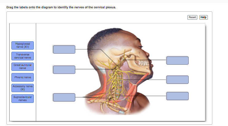
Solved Drag the labels onto the diagram to identify the ...
Description: Cervical Plexus anatomical nerve diagram, vector illustration medical scheme with human head and neck cross section. You may also like… Peripheral nervous system, medical vector illustration diagram

Nerve plexus - Wikipedia
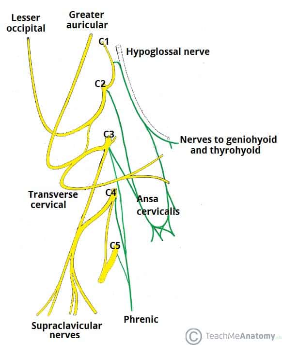
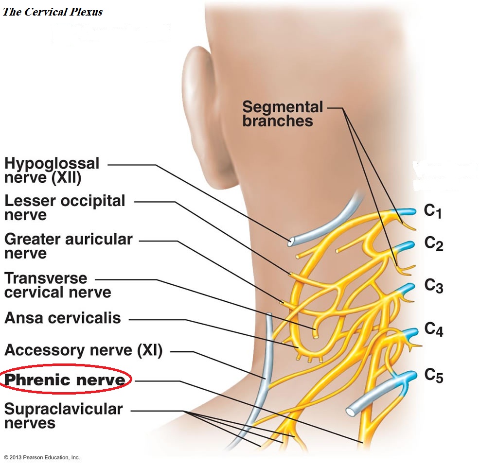
Figure 13.8 The cervical plexus. Hypoglossal nerve (XII) C 1 C 2 C 3 C 4 C 5 Segmental branches Lesser occipital nerve Greater auricular nerve Ansa cervicalis Phrenic nerve Supraclavicular nerves Accessory nerve (XI) Transverse cervical nerve Ventral rami: Ventral rami

Cervical Plexus Mohammed Albaqer Mawash Mohammed Shafik A ...
Diagram of the cervical plexus as it emerges from the cervical roots of C2-4. Note the close relationship between the cervical plexus and the phrenic nerve, which is usually affected by a deep cervical plexus block. Fig. 13.2. Branches of the cervical plexus in situ.

Cervical plexus - UpToDate
The cervical plexus is a plexus of the anterior rami of the first four cervical spinal nerves which arise from C1 to C5 cervical segment in the neck.

cervical and brachial plexus
A nerve plexus is a network of nerves that innervate the same region of the body. They are typically named after the regions of the body they innervate also, such as the cervical, brachial, lumbar ...

Local anaesthesia for carotid endarterectomy - Continuing ...
A quick, functional drawing of the cervical plexus of nerves in the neck. Feel free to ask questions in the comments.

cervical plexus of nerves | Plexus products, Deep tissue ...
Anatomy. Cranial nerves are the 12 nerves of the peripheral nervous system that emerge from the foramina and fissures of the cranium.Their numerical order (1-12) is determined by their skull exit location (rostral to caudal). All cranial nerves originate from nuclei in the brain.Two originate from the forebrain (Olfactory and Optic), one has a nucleus in the spinal cord (Accessory) while the ...

Surface markings for deep cervical plexus block. | Download ...
Cervical plexus (Gray's illustrations) Case contributed by Assoc Prof Craig Hacking . Diagnosis not applicable. Diagnosis not applicable. Edit case Share Add to. Report problem with Case.
The Cervical Plexus - Spinal nerves - Branches - TeachMeAnatomy
The cervical plexus forms from the ventral rami of C1 to C4. It is known to anastomose with the facial nerve, hypoglossal nerve, spinal accessory nerve, vagus nerve, and the sympathetic trunk. It lies anteromedial to the scalenes, but is deep to the sternocleidomastoid,[7]and gives rise to the motor and sensory branches: Motor

Cervical Plexus | Draw it to Know it
The cervical plexus is a conglomeration of cervical nerves formed by the anterior/ventral rami of spinal nerves C1-C4 (a.k.a. 1st-4th cervical nerves).These are the roots (limbs) of the cervical plexus. However, most authors include the fifth cervical nerve (i.e. the anterior ramus of spinal nerve C5) to the plexus owing to its contribution to the formation of one of the motor branches of the ...
Cervical Plexus - Physiopedia
Illustration about Cervical Plexus anatomical nerve diagram, vector illustration medical scheme with human head and neck cross section. Illustration of health, head, cord - 111598848

Cervical Plexus - Physiopedia
Nerves of the Cervical Plexus - Labeling Diagram. Higher Resolution PDF for Printing. Click Here. Use Teacher Login to show answer keys or other teacher-only items. Citing Research References. When you research information you must cite the reference. Citing for websites is different from citing from books, magazines and periodicals.

Cervical plexus - Gross Anatomy Flashcards | Draw it to Know it
Start studying Cervical Plexus. Learn vocabulary, terms, and more with flashcards, games, and other study tools.

Anatomy of cervical plexus. | Download Scientific Diagram
The cervical plexus is a plexus of the anterior rami of the first four cervical spinal nerves which arise from C1 to C4 cervical segment in the neck. They are located laterally to the transverse processes between prevertebral muscles from the medial side and vertebral (m. scalenus, m. levator scapulae, m. splenius cervicis) from lateral side. There is anastomosis with accessory nerve ...
The cervical plexus: anatomy and ultrasound guided blocks
Cervical Plexus - Color Diagram. High Resolution Poster. Click Here. Citing Research References. When you research information you must cite the reference. Citing for websites is different from citing from books, magazines and periodicals. The style of citing shown here is from the MLA Style Citations (Modern Language Association).

Drag and drop the nerves below into the bins of the plexus ...
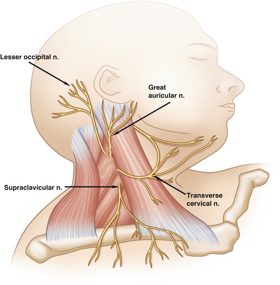
The superficial branches are the great auricular nerve, lesser occipital nerve, transverse cervical, suprasternal, and supraclavicular nerves. The deep branches are the phrenic, communicantes cervicales, communicating, and muscular. Ansa Cervicalis (C1-3) Superior (C1-2) & inferior (C2-3) roots form loop. Sensory: None.

An Overview of the Cervical Plexus - ScienceDirect
cervical plexus. I am formed by the ventral rami of the first four cervical nerves who am I. again you are the cervical plexus. most branches are_____nerves that supply only the skin. cutaneous. I transmit sensory impulses from the skin of the neck the ear area the back of the head and the shoulder who am I.

Nerves of the Cervical Plexus - Labeling Diagram
ADVERTISEMENTS: In this article we will discuss about the dissection of rat. Also learn about:- 1. Dissection of Alimentary System 2. Dissection of Circulatory System 3. Dissection of Venous System 4. The Arterial System 5. Dissection of Cranial Nerves 6. Dissection of Brain 7. Dissection of Neck Region 8. Dissection of Urinogenital System 9. The […]

Cervical Plexus | Clinical Gate
Nerves of the Cervical Plexus - Labeling Diagram. Nerves of the Lower Limb - Labeling Diagram. Nerves of the Lumbar Plexus - Labeling Diagram. Nerves of the Sacral Plexus - Labeling Diagram. Nerves of the Upper Limb - Labeling Diagram. Nervous System - Label the Parts.
Cervical plexus | Psychology Wiki | Fandom
Cervical Plexus - Color Diagram
Cervical Plexus Blocks | SpringerLink

Cervical Plexus Facts | Location, Formation, Branches ...
Online Atlas of Electromyography
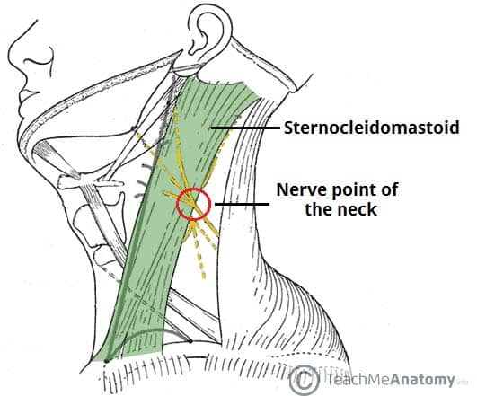
The Cervical Plexus - Spinal nerves - Branches - TeachMeAnatomy

Figure, Cervical Plexus. Purchased From Shutterstock ...

The cervical plexus - ScienceDirect

Cervical plexus, brachial plexus, lumbar plexus, sacral p...

Cervical Plexus Flashcards | Quizlet

Cervical plexus (diagram) | Image | Radiopaedia.org

Deep cervical plexus block. | Download Scientific Diagram

How to Biology & Anatomy: Cervical plexus nerves

CERVICAL PLEXUS
Do the phrenic nerves arise from the cervical plexuses, the ...

The cervical plexus, ansa cervicalis and hypoglossal nerve ...
The Cervical Plexus - Spinal nerves - Branches - TeachMeAnatomy

Cervical plexus - Wikipedia
Cervical plexus: Anatomy, branches, course, innervation | Kenhub

Deep cervical plexus block. | Download Scientific Diagram
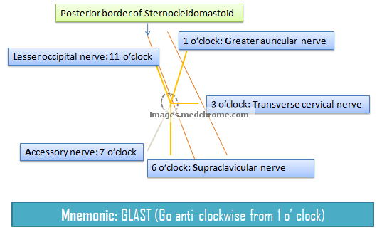
Superficial Cervical Plexus Block : Mnemonic | Epomedicine


Komentar
Posting Komentar