42 hair diagram labeled
Instructor, The Hair Diagram Online Education. Tamika Gibson is a recognized beauty influencer and educator, beautifying the world one wig at a time. Born and raised in Flint Michigan, Tamika Gibson thought she was going to be a school teacher. While studying elementary education at Western Michigan University, Tamika looked around the ... Draw A Neat Labelled Diagram Of Root Tip Showing Root Hair Zone. The Figure Given Below Is A Diagrammatic Representation Of A Part Of The Cross Section Of The Root In The Root Hair Zone Sarthaks Econnect Largest Online Education Community. Solved Explain With Well Labelled Diagram Different Regions Of Root And Also Explain Structure Of Root Hair.
The vulva (plural: vulvas or vulvae; derived from Latin for wrapper or covering) consists of the external female sex organs.The vulva includes the mons pubis (or mons veneris), labia majora, labia minora, clitoris, vestibular bulbs, vulval vestibule, urinary meatus, the vaginal opening, hymen, and Bartholin's and Skene's vestibular glands.The urinary meatus is also included as it opens into ...

Hair diagram labeled
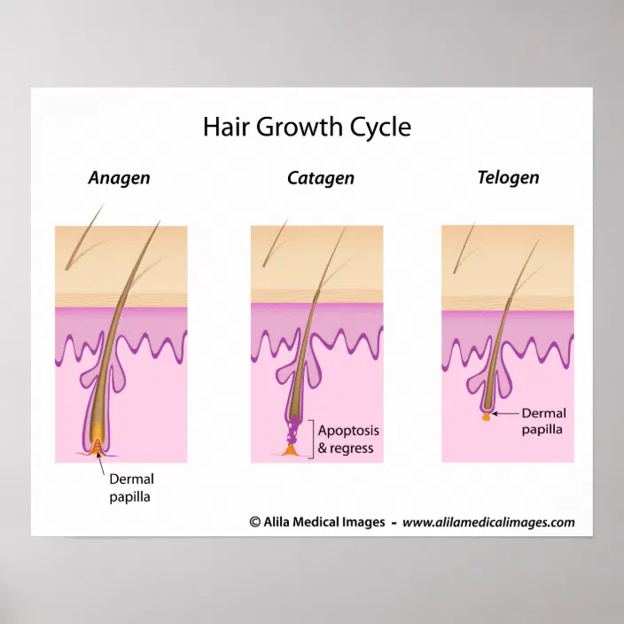
The Growth Cycle. The hair on your scalp grows about a half a millimeter a day. The individual hairs are always in one of three stages of growth: anagen, catagen, and telogen. Stage 1: The anagen phase is the growth phase of the hair. Most hair spends three to four years in this stage. Find Human Hair Follicle Labeled stock images in HD and millions of other royalty-free stock photos, illustrations and vectors in the Shutterstock collection. Thousands of new, high-quality pictures added every day. The part where both the blades meet is called the joint. Some scissors also have a logo that indicates the name of the shear manufacturer, the model number, steel used, and size. Speaking of size, this pertains to the total length of the scissors. It is measured from the points to the edge of the finger bows.
Hair diagram labeled. Hair is a keratinous filament growing out of the epidermis. It is primarily made of dead, keratinized cells. Strands of hair originate in an epidermal penetration of the dermis called the hair follicle. The hair shaft is the part of the hair not anchored to the follicle, and much of this is ... Thick and thin skin histology labeled diagram. I would like to show you the different histological features from both thick and thin skin histology slides with a labeled diagram. I hope these skin microscope slide labeled diagrams might help you to identify and learn all the structures. The below figure shows a root hair: (i) Label the parts 1 to 4. (ii) What is the role of part 4? (iii) Why is the root hair one-celled? (iv) What will happen to the root hair if some fertilizer is added to the soil near the root hair? The lines are labeled from left to right as Perch, Frog, Pigeon, Rats, and Human. A text box below the branching tree diagram is labeled Derived Shared Characteristics. In the box it says from left to right, Terrestrial during all stages, Jaws, Walking on two legs, Mammary glands and hair, and Four limbs.
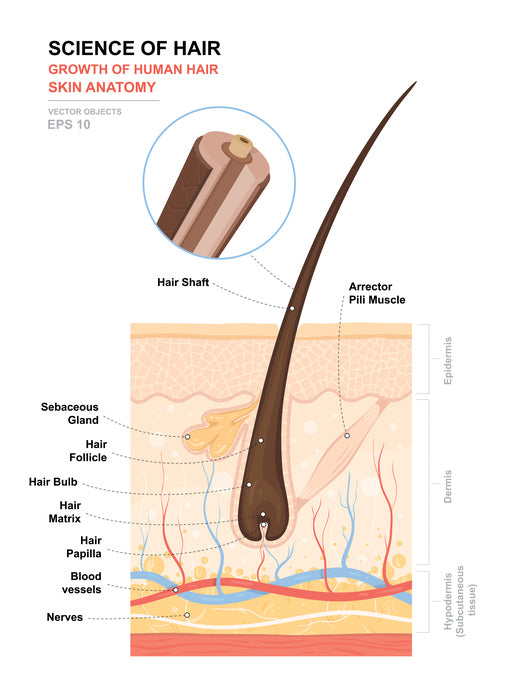
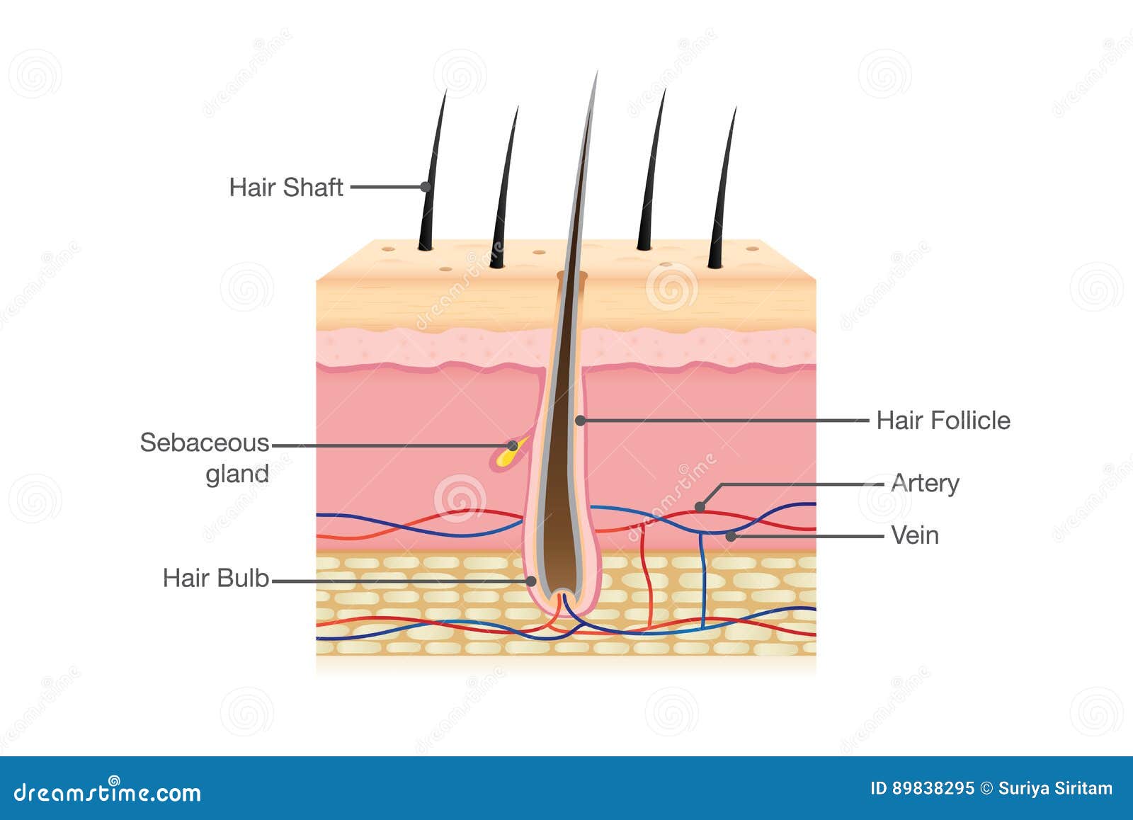
Illustration of human Hair Anatomy. Diagram of a hair follicle and cross section of the skin layers vector art, clipart and stock vectors. Image 69363580. Dec 27, 2021 · Labeled Hair Follicle Diagram Labeled Hair Follicle Diagram 133231 Layers Of The Skin Skin Anatomy Anatomy And Physiology Integumentary System. Base Of Hair Follicle Human Body Anatomy Medical Anatomy Chemistry Education. Shows A Root Hair Cell The Water Can Pass Through The Thin Cell Wall Of The Root Hair Biology Revision Photosynthesis Cell Wall. Cardiovascular system - diagram. Small veins, called venules, leave from capillaries and gradually increase their lumen on the way to the heart to end as veins.There is a certain histological difference between arteries and veins, but their main functional difference reflects the direction in which they conduct blood: the arteries convey blood from the heart to the periphery, whereas the veins ... Hair helps to protect the body from UV radiation by preventing sunlight from striking the skin. Hair also insulates the body by trapping warm air around the skin. The structure of hair can be broken down into 3 major parts: the follicle, root, and shaft. The hair follicle is a depression of epidermal cells deep into the dermis.
The bird feather diagram below displays the different parts of the bird feather. It is labeled with the calamus (quill), rachis (shaft), barbs, and vane. A diagram of the anatomy of a feather. The structure labeled as Q which is a large structure in the root hair cell covering most of its area represents the vacuole of the root hair cell, and the structure labeled as R represents the cell wall of the root hair cell that is an outer covering of the root hair cell. Honey Bee Anatomy. Bees Have 3 Main Body Parts. Honey Bee Stingers. Honey Bee Crop - Honey Stomach. FAQs About Honey Bee Anatomy. Though small in size, this insect's body is a complex arrangement of specialized structures. All of the parts of a honey bee work together to make survival possible. Apr 1, 2014 - This Pin was discovered by Lindsay Catania. Discover (and save!) your own Pins on Pinterest
Label The Features Of The Hair Follicle Diagram | Quizlet
39,281 hair structure stock photos, vectors, and illustrations are available royalty-free. See hair structure stock video clips. of 393. hormonal skin epidermis 3d hair folicule hair follicle scalp young skin old skin hair anatomy hair section layer of human skin scalp follicles fat tissue in section. Try these curated collections.

The Structure Of The Hair | Chic Girl Online
May 3, 2017 - Hair is one of the characteristic features of mammals and has various functions such as protection against external factors; producing sebum, apocrine sweat and pheromones; impact on social and sexual interactions; thermoregulation and being a resource for stem cells.
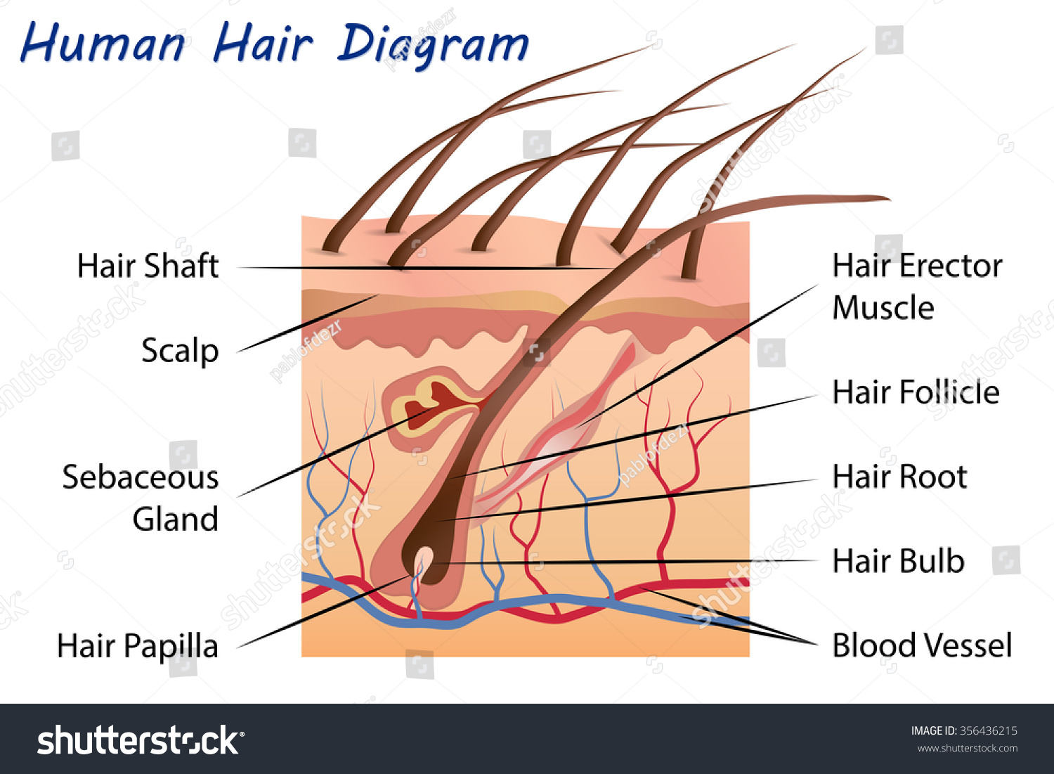
Human Hair Diagram - Royalty Free Stock Vector 356436215 ...
Your hair is made of two parts - the hair follicle and the hair shaft. Hair follicle anchors or holds the hair into the scalp. It has the following parts : 1. Bulb. The bulb is found at the root of your hair where the protein cells (keratin) grow to make hair. 2. Papilla. The papilla provides blood supply to the hair follicles for healthy hair. 3.

Human Skin Cell Crosssection Of The Structure Labeled Hair ...
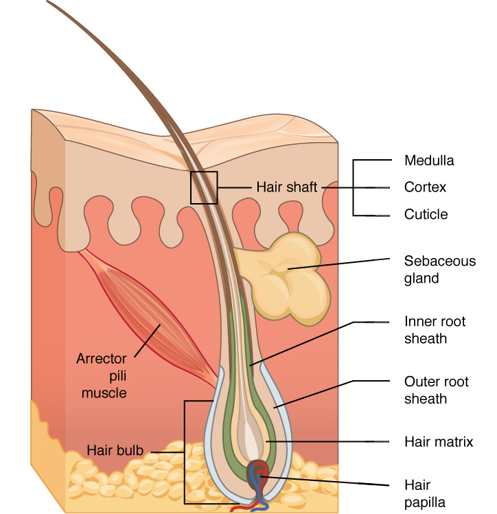
Figure 1. Hair Diagram A hair grows from the papilla and with the exception of that point of generation is made up of dead, cornified cells. It consists of a shaft that projects above the skin, and a root that is imbedded in the skin. Figure 2 diagrams how the lower end of the root expands to form the root bulb.
(118).jpg)
A Human Body Skin-structure Quiz! - ProProfs Quiz
Anatomy of hair - download this royalty free Vector in seconds. No membership needed.

Hair Anatomy Structure Diagram Vector Illustration Stock ...
The muzzle is the part of the horse's head that includes the area of the mouth, nostrils, chin, lips, and front of the nose. The muzzle is very mobile and sensitive. Whiskers help the horse sense things close to its nose and the skin is almost hairless. Beneath the skin is cartilage. Continue to 2 of 29 below.

Illustration Of Anatomy Of Hair With Label On White ...
Normal hair follicle and skin with Alopeci. A. human skin. Vector illustration · Acne types. Pimple skin diseases anatomy medical vector diagram. Illustration of follicle and pimple, medicine anatomy, papule and pustule · Labeled Skin and hair anatomy. Detailed medical illustration

diagram of a hair follicle in a cross section of skin | Piel ...
Integumentary system quiz and answers. One of the best ways to start learning about a new system, organ or region is with a labeled diagram showing you all of the main structures found within it. Not only will this introduce you to several new structures together, it will also give you an overview of the relations between them.

VTCT - Functions and Structure of the Skin
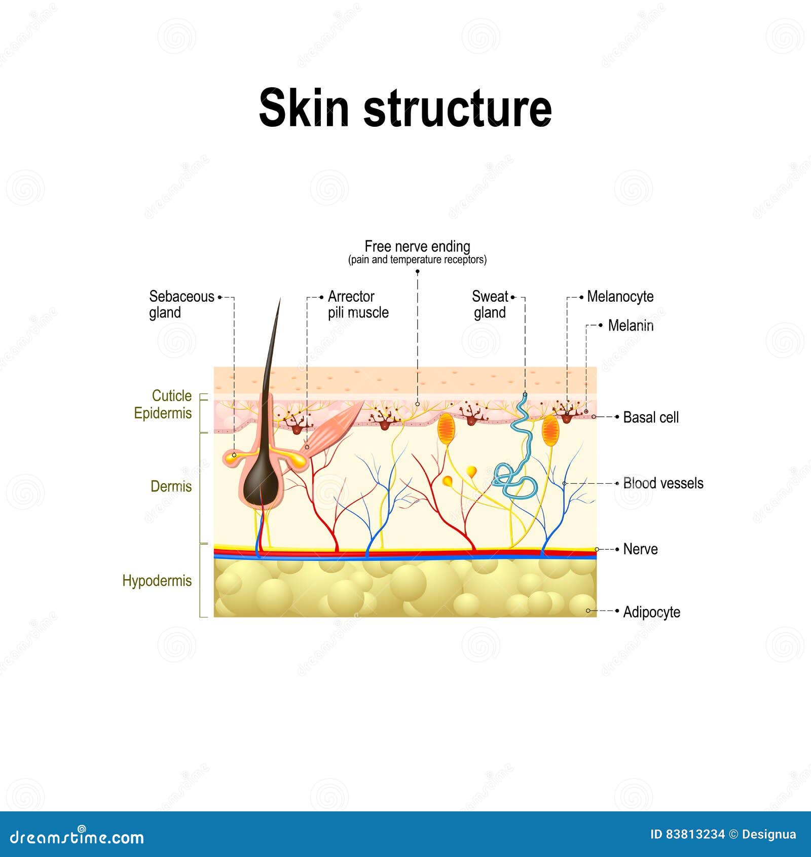
The dermis is a connective tissue layer sandwiched between the epidermis and subcutaneous tissue. The dermis is a fibrous structure composed of collagen, elastic tissue, and other extracellular components that includes vasculature, nerve endings, hair follicles, and glands. The role of the dermis is to support and protect the skin and deeper layers, assist in thermoregulation, and aid in ...

Human hair diagram piece Royalty Free Vector Image
Start studying Labeling of Hair Diagram Structures. Learn vocabulary, terms, and more with flashcards, games, and other study tools. Scheduled maintenance: Saturday, March 6 from 3-4 PM PST
Human hair diagram Images, Stock Photos & Vectors | Shutterstock
Test your knowledge on this science quiz to see how you do and compare your score to. Basic hair structure explained. Learn how all parts of the hair shaft function ( cuticle, medulla and cortex layers) using the hair shaft and hair follicle diagrams. Human hair follicle, labeled - buy this illustration on Shutterstock & find other images.

Labeled Skin Diagrams | Health Pictures | Skin anatomy, Skin ...
Oh no! Pinterest doesn't work unless you turn on JavaScript
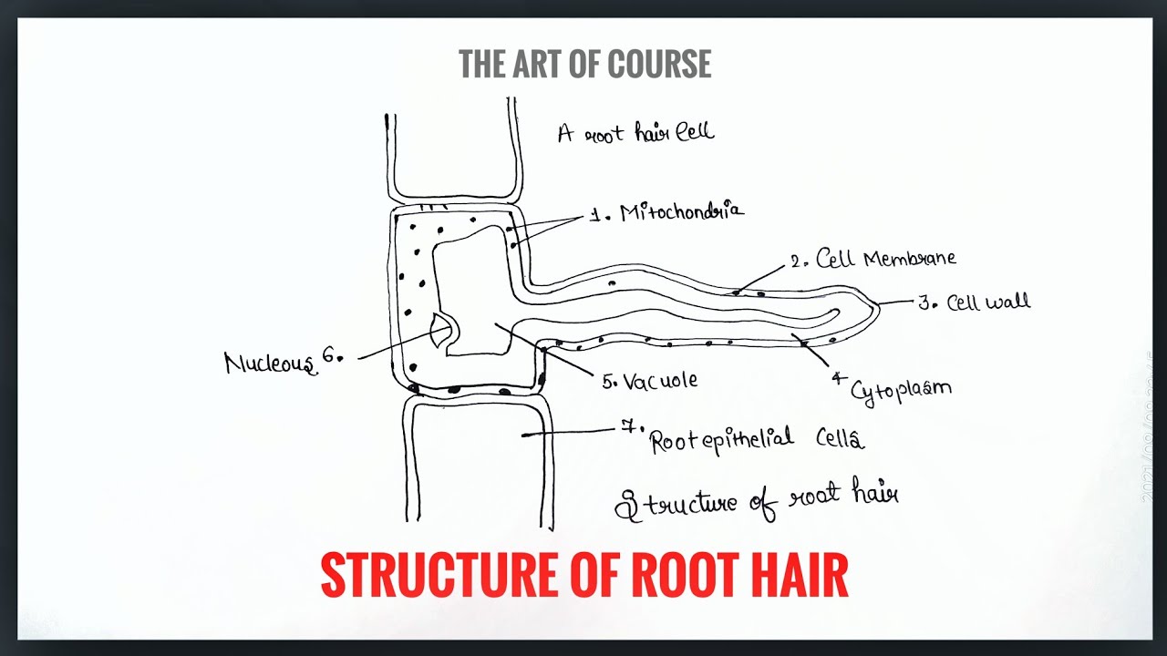
Structure of Root Hair Diagram with labelling ...
Scalp and hair histology Author: Rachel Baxter BSc, MSc • Reviewer: Francesca Salvador MSc Last reviewed: September 30, 2021 Reading time: 11 minutes Covering the surface of your head, the scalp, extends from the top of your forehead across to the epicranial aponeurosis of the head.Laterally, it reaches down to the external auditory meatus and zygomatic arch (cheekbone of the skull).
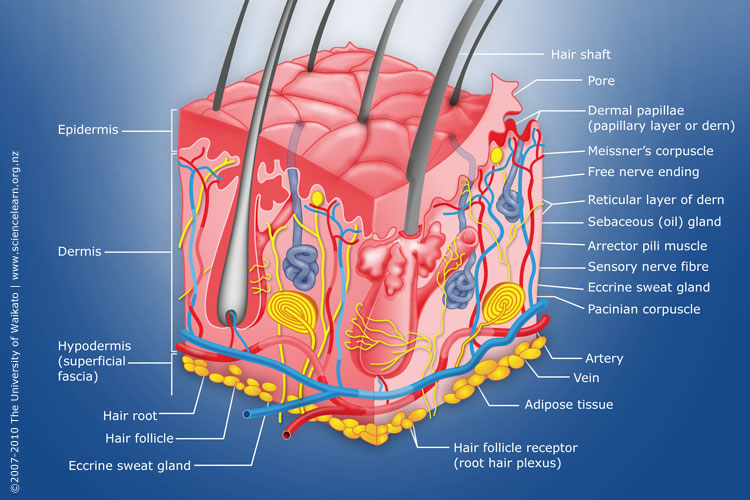
Diagram of human skin structure — Science Learning Hub
Start studying Hair structure diagram. Learn vocabulary, terms, and more with flashcards, games, and other study tools.
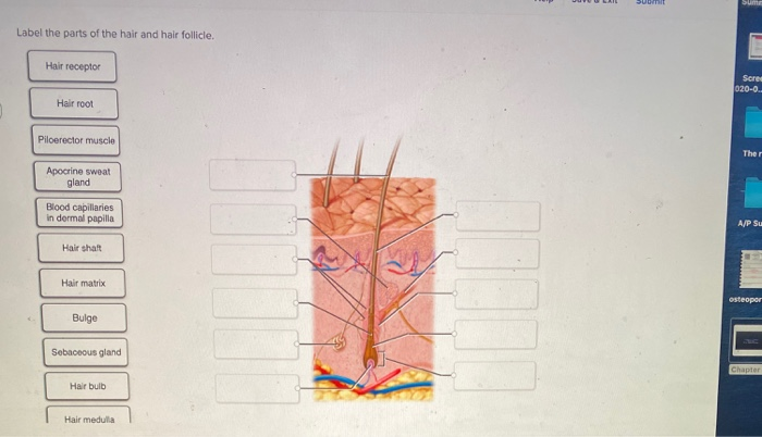
Solved Label the parts of the hair and hair follicle. Hair ...
Set of Human Sensory Organs: Anatomy of Five Sensory Organs, Eyes, Ear, Skin, Nose and Tongue. Sensory Organs provide us with data for perception, and it is the physiological capacity of all living organisms. Five sense organs are equipped in the human body. Those organs provide us with first-hand information about our external or internal world.
Hair | Biology for Majors II
Skin is the largest organ in the body and covers the body's entire external surface. It is made up of three layers, the epidermis, dermis, and the hypodermis, all three of which vary significantly in their anatomy and function. The skin's structure is made up of an intricate network which serves as the body's initial barrier against pathogens, UV light, and chemicals, and mechanical injury.

Human Hair Anatomy. Diagram Of A Hair Follicle And Cross ...
A heart diagram labeled will provide plenty of information about the structure of your heart, including the wall of your heart. The wall of the heart has three different layers, such as the Myocardium, the Epicardium, and the Endocardium. Here's more about these three layers. Epicardium

Anatomy of a Hair Follicle Quiz/Worksheet by Everything ...
Rich in Diversity, Committed to Excellence · On January 31, 2022, CASD is hosting Woodlyn Pharmacy in the CASH auditorium hallway. The hours will be from 4:00pm to 7:30pm, for all Coatesville Area residents ages 5 and older. You must register using these links below for both your booster or ...
Hair follicle
September 3, 2017 - Complexion, Physiology, Integumentary System, Medical, Healthy, Beauty, Cosmetic, Makeup, Treatment Stock vector 165128754 ⬇ Download from Depositphotos ⚡ Millions of royalty-free vector images & illustrations.
Label a diagram of the skin - Mrs. Sanborn's Science Class
Each hair on your body grows from a hair follicle, a tiny, saclike hole in your skin. At the bottom of each follicle is a cluster of special cells that reproduce to make new hair cells. The new cells that are produced are added on at the root of the hair, causing the hair to grow longer · ...
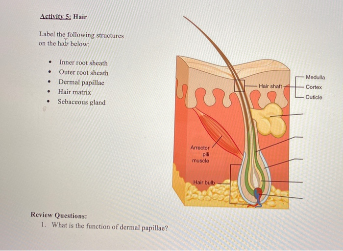
Solved Activity 5: Hair Label the following structures on ...
Aug 15, 2018 · Labeled diagram of hair follicle quizlet together with story along with page along with lesson 01 20 e2 80 93 20skin 20structure 20and 20function together with chapter 5 integumentary system flash cards together with labeled diagram of hair in addition anatomy and physiology of healthy skin as well as image of a cheek cell schematic.Structure ...

Drawing shows how hair follicle was divided into three parts ...
Drag and drop the pins to their correct place on the image.
Understanding the Anatomy of Hair - Toppik Blog
Labeled diagram of the external genital organs that make up the vulva. Mons Pubis, Labia Majora, and Labia Minora ... The hair pattern on the mon pubis varies by racial and genetic factors. The ...
Hair growth cycle labeled version diagram. poster | Zazzle.com
July 23, 2021 - © 2014 WebMD, LLC. All rights reserved · Hair is simple in structure, but has important functions in social functioning. Hair is made of a tough protein called keratin. A hair follicle anchors each hair into the skin. The hair bulb forms the base of the hair follicle.
Hair Anatomy: Overview, Microanatomy of Anagen Phase Hair ...
Figure 1. Hair follicles originate in the epidermis and have many different parts · Hair is a keratinous filament growing out of the epidermis. It is primarily made of dead, keratinized cells. Strands of hair originate in an epidermal penetration of the dermis called the hair follicle.

Skin anatomy realistic background with layers and labeled parts vector illustration
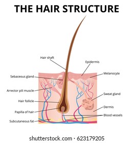
The hair is made up of 95% keratin, a fibrous, helicoidal protein that forms part of the skin and all its appendages (body hair, nails, etc.)...

Why Does Hair Turn Gray? - Kristin Moon Science
Start studying Anatomy 2017- Unit 2 Label Hair Diagram. Learn vocabulary, terms, and more with flashcards, games, and other study tools.

Intro to the Integumentary System Quiz - Quizizz
Cilia and Flagella are tiny hair-like projections from the cell made of microtubules and covered by the plasma membrane. ... Structure, Parts, Functions, Labeled Diagram, Worksheet; Animal Cell- Definition, Organelles, Structure, Parts, Functions, Labeled Diagram, Worksheet; Amazing 27 things under the microscope (diagrams and descriptions)

Medical Education Chart of Biology for Hair Diagram Stock ...
Vagina: The vagina is a muscular canal that connects the cervix and the uterus, leading to the outside of the body. Parts of the vagina are rich in collagen and elastin, which give it the ability to expand during sexual stimulation and childbirth. Cervix: The cervix is the lower part of the uterus that separates the lower uterus and the vagina and may play a role in lubrication.
Human Hair Diagram Stock Illustrations – 1,300 Human Hair ...
human hair diagram images. 2,841 human hair diagram stock photos, vectors, and illustrations are available royalty-free. See human hair diagram stock video clips. of 29. human skin structure layer of human skin hair diagram dermatology skin cross section hair follicle vector structure of the skin structura hair old skin anatomy hair ...
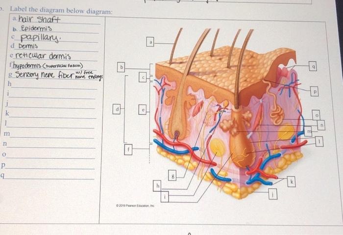
Solved a Label the diagram below diagram: a hair shaft b ...
In this tutorial, we have described Stem Anatomy (Monocot and Dicot Stem Cross Section). DICOT STEM CROSS SECTION For studying the internal structure of a typical dicot stem, the stem cross section of a young Sunflower or Cucurbita is taken. A thin transverse section of the stem reveals the following structures under the microscope: 1. Epidermis It forms a single and the outermost layer of the ...

Forensics: History of Hair/Fiber Analysis and Hair Diagram
Hair Growth in Puberty. 4 / 12. After breasts and testicles start growing, body hair will start to grow in and become thicker. For both boys and girls, new hair will start growing in the armpits ...

Vascular plant biological structure labeled diagram, vector illustration art print poster
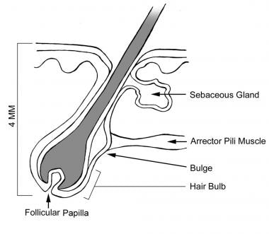
the hair follicle (important to Hair care and maintenance) the hair shaft. (important to hair grooming and dressing) (1) HAIR FOLLICLE. The hair follicle is the point from which the hair grows. It is a tiny cup-shaped pit buried in the fat of the scalp. The terminal part of the hair follicle seated within the skin is called a hair bulb.
Human Hair Diagram Stock Illustrations – 1,300 Human Hair ...
Figure 82. Hair Diagram. A hair grows from the papilla and, with the exception of that point of generation, is made up of dead, cornified cells. It consists of a shaft that projects above the skin ...

The Human Hair Anatomical Chart
ROOT ANATOMY: DICOT ROOT CROSS SECTION Dicot Root Diagram Reveals Internal Structure of Dicot Root. A thin transverse section of the young dicot root of Gram, Sunflower or Pea reveals the following structures under the microscope: 1. Epiblema or Piliferous Layer. This is the outermost layer of root-derived from the protoderm of the apical meristem.
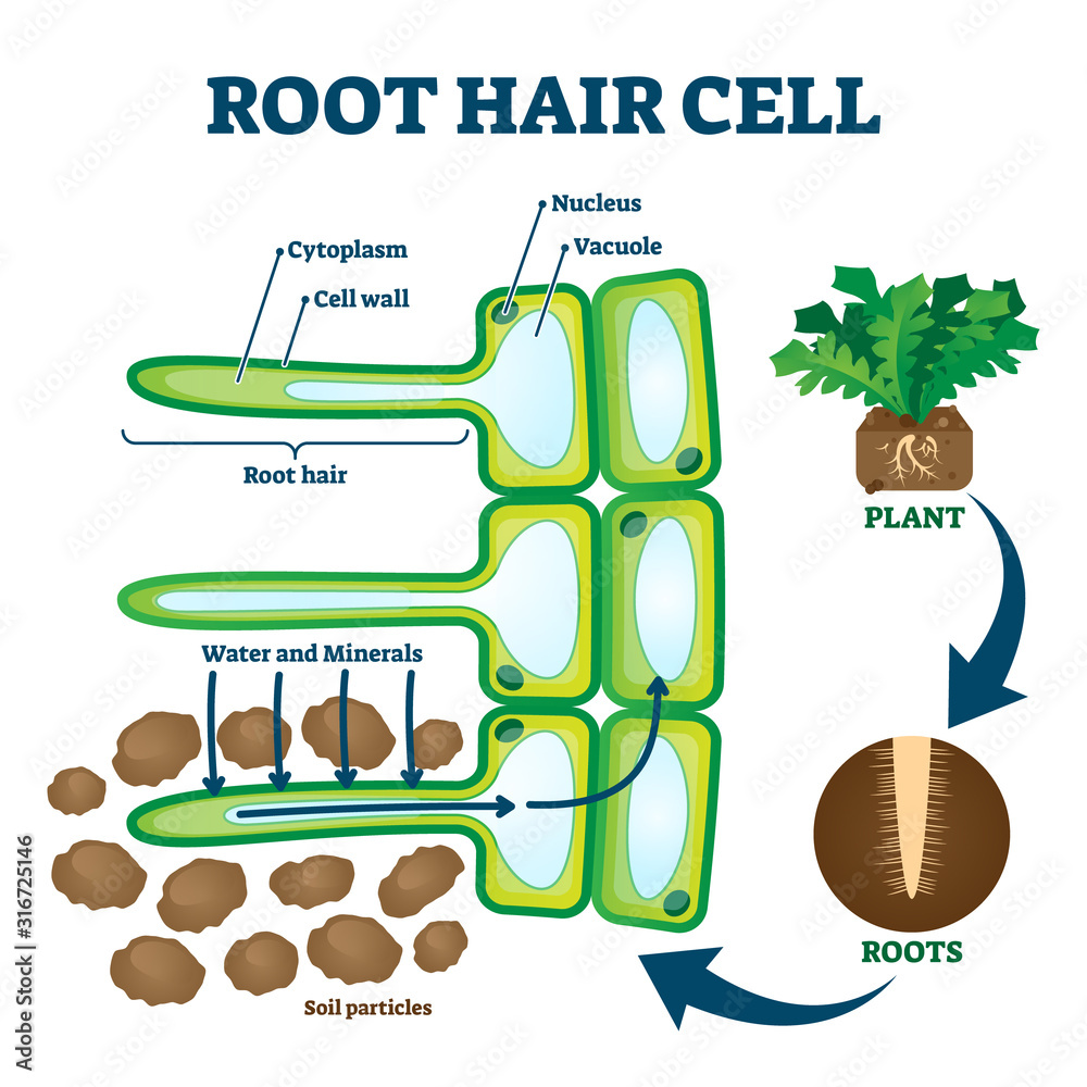
Root hair cell collecting mineral nutrients and water from ...
Download 733 Diagram Hair Structure Stock Illustrations, Vectors & Clipart for FREE or amazingly low rates! New users enjoy 60% OFF. 177,637,203 stock photos online.

33 Label The Structures Of The Hair Follicle. - Labels Design ...
Yellowing of the hair due to sunlight. Grey hair. Breakage of the hair in mid-shaft. Male pattern balding. NEXT>. 4. Hair goes through resting, growing and falling out phases. These phases are known as Anagen, Catagen and which other name? Foragen.

label the hair follicle model - Brainly.com
Integumentary System definition. The integumentary system is a system comprised of organs that are the outermost protective covering of the animal body, the skin, and its various derivatives. The integumentary system protects against many threats such as infection, desiccation, abrasion, chemical assault, and radiation damage.
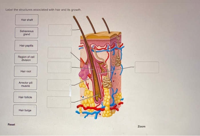
Solved Label the structures associated with hair and its ...
October 17, 2017 - Herpes simplex virus infection, labeled diagram. Herpes simplex virus (HSV-1) latent (in trigeminal nerve ganglion) and active infection (cold sores), labeled drawing. Human skin anatomy, labeled. Structure of human skin, epidermis, dermis and hypodermis layers with hair follicle, sebaceous ...

Anatomy Of Human Skin Cross Section View Labeled On White ...
The part where both the blades meet is called the joint. Some scissors also have a logo that indicates the name of the shear manufacturer, the model number, steel used, and size. Speaking of size, this pertains to the total length of the scissors. It is measured from the points to the edge of the finger bows.
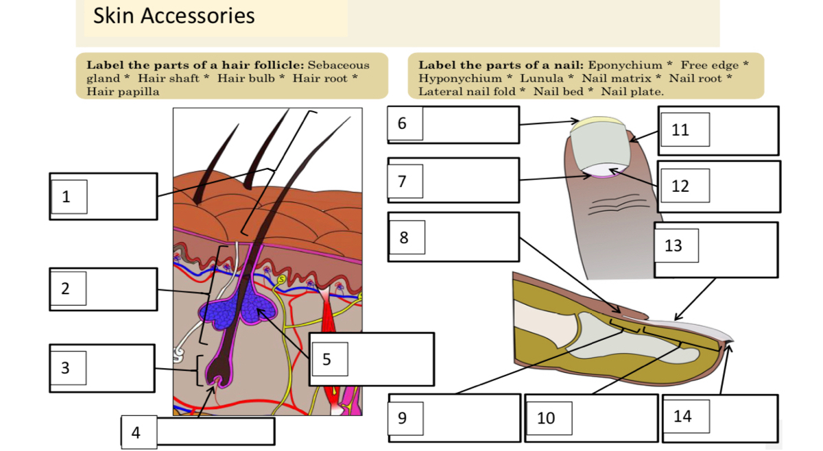
Answered: Skin Accessories Label the parts of a… | bartleby
Find Human Hair Follicle Labeled stock images in HD and millions of other royalty-free stock photos, illustrations and vectors in the Shutterstock collection. Thousands of new, high-quality pictures added every day.
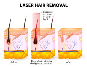
Laser Hair Removal in Plano, TX Offers Incredible Benefits
The Growth Cycle. The hair on your scalp grows about a half a millimeter a day. The individual hairs are always in one of three stages of growth: anagen, catagen, and telogen. Stage 1: The anagen phase is the growth phase of the hair. Most hair spends three to four years in this stage.


Komentar
Posting Komentar