41 microscope ray diagram
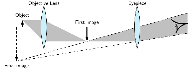
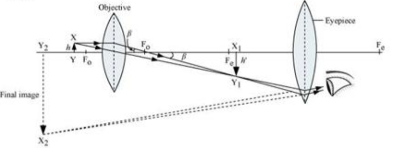
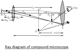
Geometrical Construction of Ray Diagrams Geometrical Construction of Ray Diagrams A popular method of representing a train of propagating light waves involves the application of geometrical optics to determine the size and location of images formed by a lens or multi-lens system. This tutorial explores how two representative light rays can establish the parameters of an imaging scenario. Which ray diagram is correct for a Compound microscope ... Here are two ray diagrams for compound microscope, the first one proposed by the book, and the second one recommended by the teacher: In the first image, the light rays form a real image A'B', which becomes the virtual object for the eyepiece. See, the original rays are carried forward to the eyepiece, which then form a virtual image, A"B".
Transmission electron microscopy(TEM) | Principle, Ray ... In this animated flip learning resource, you will study the following important concepts of phase contrast microscopy: 1. Definition of Transmission electron...

Microscope ray diagram
PDF Chapter 4 Optics - UNC School of Medicine The rays are being focused to a focal pointat the right on the lens axis. The distance from the center of the lens to the focal point is the lenses focal length. A plane drawn perpendicular to the lens axis at the focal point is the focal plane. A ray diagramlike this can be interpreted in both directions. Types of Microscopes: Definition, Working Principle ... Stereo Microscope Diagram Principle of Stereo Microscope A stereo microscope works on the reflected light from the sample. The magnification of the microscope takes place at low power and hence, it is suitable for magnifying the opaque objects. It is suitable for thick and solid samples because it uses light reflected from the sample. optics - Ray diagram of focussing on a compound microscope ... Here is the ray diagram of a compound microscope. So, when we are focussing, we move the objective lens which tweaks the image distance. My doubt is that, shouldn't the image be seen clearly, wheresoever the first real image forms, if within Fe (Focus of the eyepiece lens). Then, that would make it a range instead of a single point (shouldn't ...
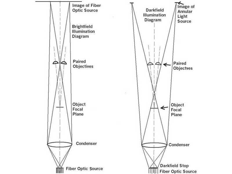
Microscope ray diagram. Brightfield Microscope (Compound Light Microscope ... Brightfield Microscope Definition. Brightfield Microscope is also known as the Compound Light Microscope. It is an optical microscope that uses light rays to produce a dark image against a bright background. It is the standard microscope that is used in Biology, Cellular Biology, and Microbiological Laboratory studies. Simple microscope - Fun Science A simple microscope works on the principle that when a tiny object is placed within its focus, a virtual, erect and magnified image of the object is formed at the least distance of distinct vision from the eye held close to the lens. Working of Simple Microscope. The ray diagram to show the working of simple microscope is shown in figure. PDF LM RAY DIAGRAMS Symmetrical Ray Diagrams of the Optical ... Such a ray diagram was produced in 1998 for the case of a light microscope with finite (i.e. 160 mm) tube length-corrected objectives [3]. Today, most new microscopes are designed with infinity-corrected optical systems, so a new set of ray diagrams is now called for. In this article we now present a set of sym-metrical ray diagrams of the ... The Working of Dark-field Microscope with Ray Diagram ... ADVERTISEMENTS: Read this article to learn about the working of dark-field microscope with ray diagram! Working Principle: In a dark-field microscope, the object is brilliantly illuminated against a dark background (Figure 4.10). This is accomplished by equipping a light microscope with a special kind of condenser. ADVERTISEMENTS: It is a condenser with a dark-field stop, […]
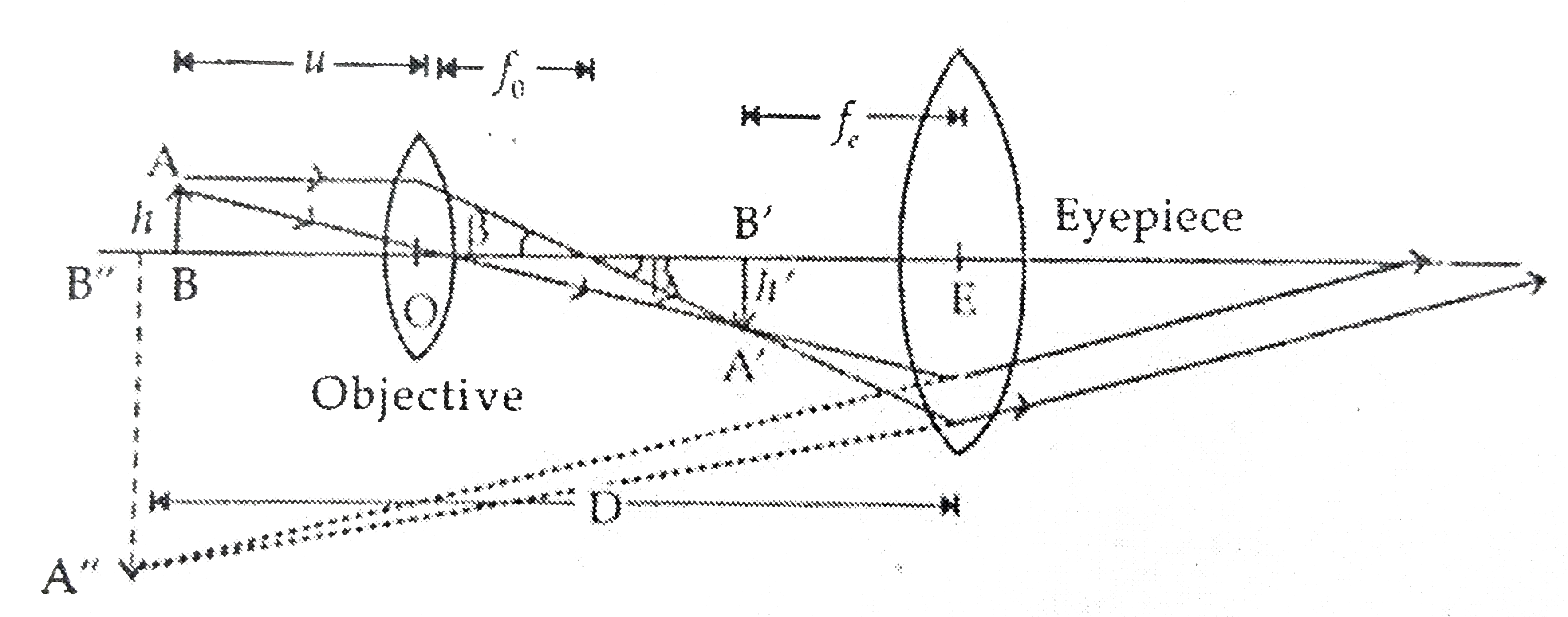
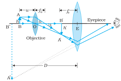
Draw a ray diagram of compound microscope, when final ... Draw a ray diagram of compound microscope, when final image is formed at the minimum distance of distinct vision. Easy Solution Verified by Toppr It consist of two convex lenses, one objective of very small focal length with short aperture. And one Eyepiece with moderate focal length and large aperture. Simple Microscope - Definition, Types, Working Principle ... A simple microscope consists of a convex lens of a short focal length. The below figure shows the ray diagram which subsequently forms the image of an object (or we can say a source of light). (Image will be Updated soon) F is the focal length of the lens. An object is placed between the focal length and the centre of the curvature. (a) Draw a labelled ray diagram of a compound microscope ... (a) Labelled diagram of compound microscope. The objective lens form image A' B' near the first focal point of eyepiece. (b) Angular magnification of objective lens m 0 = linear magnification h'/h. where L is the distance between second focal point of the objective and first focal point of eyepiece.If the final image A'' B'' is formed at the near point. Geometrical Construction of Ray Diagrams | Nikon's MicroscopyU Geometrical Construction of Ray Diagrams A popular method of representing a train of propagating light waves involves the application of geometrical optics to determine the size and location of images formed by a lens or multi-lens system. This tutorial explores how two representative light rays can establish the parameters of an imaging scenario.
Draw a labelled ray diagram of a compound microscope and ... Draw a labelled ray diagram of a compound microscope and write an expression for its magnifying power. The focal length of the objective and eye-lens of a compound microscope are 2cm, 6.25cm respectively. The distance between the lenses is 15cm. Transmission Electron Microscopy (TEM) Figure 2 - A ray diagram for the diffraction mechanism in TEM Imaging The beam of electrons from the electron gun is focused into a small, thin, coherent beam by the use of the condenser lens. This beam is restricted by the condenser aperture, which excludes high angle electrons. Compound Microscope: Definition, Diagram, Parts, Uses ... Compound microscope is a type of optical microscope that is used for obtaining a high-resolution image. There are more than two lenses in a compound microscope. Learn about the working principle, parts and uses of a compound microscope along with a labeled diagram here. (a) Draw a labelled ray diagram of compound microscope ... (a) Draw a labelled ray diagram of compound microscope, when final image forms at the least distance of distinct vision. (b) Why is its objective of short focal length and of short aperture, compared to its eyepiece? Explain. (c) The focal length of the objective is 4 cm while that of eyepiece is 10 cm. The object is placed at a distance of 6 cm from the objective lens.

Confocal Microscopy - Microscopist.co.uk
Draw a ray diagram of compound microscope when the class ... The course of rays through a compound microscopic is shown in the given figure. Here we can see that an image is formed at the least distance of distinct vision from eyes. Here, AB ia a tiny object held perpendicular to the common principal axis, in front of the objective lens beyond its focus.
![PDF] Symmetrical Ray Diagrams of the Optical Pathways in ...](https://d3i71xaburhd42.cloudfront.net/01d8132b06db2a6255ebd5f93317d2966b1e59e0/3-Figure3-1.png)
PDF] Symmetrical Ray Diagrams of the Optical Pathways in ...
Fluorescence microscopy | Principle, Ray diagram, design ... In this animated flip learning resource, you will study the following important concepts of phase contrast microscopy: 1. Definition of fluorescence compound...
a) Draw a ray diagram showing the image formation by a ...
Ray Diagrams - Physics Classroom The description is applied to the task of drawing a ray diagram for an object located beyond the 2F point of a double convex lens. 1. Pick a point on the top of the object and draw three incident rays traveling towards the lens. Using a straight edge, accurately draw one ray so that it passes exactly through the focal point on the way to the lens.
Which ray diagram is correct for a Compound microscope ...
(a) Draw the labelled ray diagram for the formation of ... Click here👆to get an answer to your question ️ (a) Draw the labelled ray diagram for the formation of image by a compound microscope. Derive an expression for its total magnification (or magnifying power), when the final image is formed at the near point.(b) Why both objective and eyepiece of a compound microscope must have short focal lengths?Draw a ray diagram showing the image ...

Draw a Labelled Ray Diagram Showing the Formation of Image by ...
CBSE NCERT Notes Class 12 Physics Ray Optics Optical ... Class 12 Physics Ray Optics Optical Instruments. Microscope. Microscope. Microscope is an instrument that gives an enlarged image of minute object. There are 2 types of microscope:-. Simple. Compound. Simple Microscope. An instrument that gives an enlarged image of a minute object.

Dissecting/Stereo microscope | Principle, Parts, working, and ...
Darkfield Microscope- Definition, Principle, Uses, Diagram A dark field microscope is arranged so that the light source is blocked off, causing light to scatter as it hits the specimen. This is ideal for making objects with refractive values similar to the background appear bright against a dark background. When light hits an object, rays are scattered in all azimuths or directions.
Exercises, Telescopes and microscopes, By OpenStax (Page 2/2 ...
Draw a ray diagram of a compound microscope. Write the ... Ray diagram of a compound microscope.When the final image is formed at the least distance of distinct vision,For the image formed at infinity, ue = feand By making focal length of the objective small, the magnifying power can be increased.
Draw the labeled ray diagram for the formation of image by a ...
The Microscope Optical Train - Nikon's MicroscopyU In considering the action of lenses, the wave-like properties can often be ignored and light is considered to travel in straight lines often termed rays. Simple ray diagrams are sufficient to explain many important aspects of microscopy including refraction, focal length, magnification, image formation, and diaphragms.

Ray diagrams showing the important optical elements for (a ...
Physics Questions For full functionality of this site it is necessary to enable JavaScript. Here are the instructions how to enable JavaScript in your web browser.
Mic-UK: Introducing Children to the Micro-Life of Fish Lake
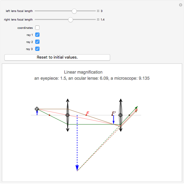
Ray Diagrams for Microscope and Telescope - Wolfram ... Ray Diagrams for Microscope and Telescope. Copied! Copying... Interact on desktop, mobile and cloud with the free Wolfram Player or other Wolfram Language products. This Demonstration shows the linear magnification for a pair of lenses such as in a microscope or telescope.

View Image
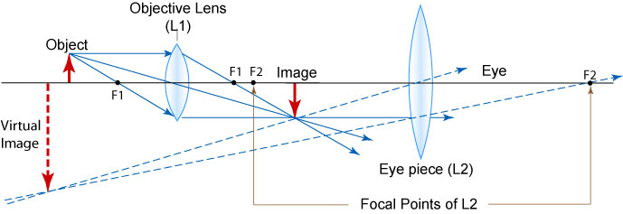
Lenses - Boston University A microscope arrangement is shown below, along with the ray diagram showing how the first lens creates a real image. This image is the object for the second lens, and the image created by the second lens is the one you'd see when you looked through the microscope. Note that the final image is virtual, and is inverted compared to the original ...

Schematic ray diagram of the Zernike phase contrast X-ray ...
optics - Ray diagram of focussing on a compound microscope ... Here is the ray diagram of a compound microscope. So, when we are focussing, we move the objective lens which tweaks the image distance. My doubt is that, shouldn't the image be seen clearly, wheresoever the first real image forms, if within Fe (Focus of the eyepiece lens). Then, that would make it a range instead of a single point (shouldn't ...

Principles of imaging with an optical microscope: (a) ray ...
Types of Microscopes: Definition, Working Principle ... Stereo Microscope Diagram Principle of Stereo Microscope A stereo microscope works on the reflected light from the sample. The magnification of the microscope takes place at low power and hence, it is suitable for magnifying the opaque objects. It is suitable for thick and solid samples because it uses light reflected from the sample.

Ray optics diagrams of the objective-based TIRF microscopy ...
PDF Chapter 4 Optics - UNC School of Medicine The rays are being focused to a focal pointat the right on the lens axis. The distance from the center of the lens to the focal point is the lenses focal length. A plane drawn perpendicular to the lens axis at the focal point is the focal plane. A ray diagramlike this can be interpreted in both directions.

optics - Ray diagram of focussing on a compound microscope ...

draw a ray diagram to show the image formation by a compound ...

Ray paths in an inverted microscope set up for K ̈hler ...
Ray Diagrams for Microscope and Telescope - Wolfram ...
Compound Microscope, Ray Diagram Mistakes. | Physics Forums

Optical Instruments: Compound Microscope and its Magnification

Optical ray tracing using Zemax © (a) and (b) shows the ray ...

Ray Diagram of a Simple Microscope | Diagram, Simple, Life ...

File:LEED Lens and Ray Diagram.png - Wikipedia
1 Light Microscopy

a Draw a ray diagram for the formation of image by a compound ...

Microscopy, optical and electron microscope with ray diagram

Draw a ray diagram of compound microscope, when final image ...

Draw the Ray diagram of image formation in simple microscope ...

7 Ray diagram for a transmission electron microscope in image ...

Compound Microscope

The Compound Microscope - ppt download
2 Ray diagram of a basic electron microscope (a) Ray diagram ...

Draw the ray diagram of image formation in case of compound ...

Answer the question number g Q (g) Draw a ray diagram to show ...
Compound Microscope, Ray Diagram Mistakes. | Physics Forums

Draw a ray diagram of compound microscope, when final image ...

Draw the ray diagram of image formation in case of compound ...
Draw a ray diagram of compound microscope when the class 12 ...
Draw the labelled ray diagram for the formation of image by a ...
Draw a ray diagram to show the image formation by a compound ...
ray diagram | cell in life


Komentar
Posting Komentar