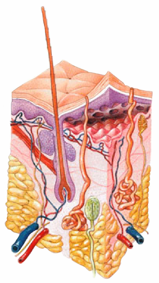
39 skin diagram blank
PDF SELF EXAMINATION HOW TO USE THE BODY MAP BODY ... - Skcin Draw a line from each dot and record the date, colour, size and shape of each mole identified and make a note of any patches of skin that appear abnormal and how they look and feel, like the example provided (right). Skin Diagrams Teaching Resources | Teachers Pay Teachers Good diagrams are a must for teaching human body systems! Enhance your study of the Integumentary System with this clear diagram of the Human Skin in cross-section in both print (black & white) and digital (color) formats. Also includes EDITABLE matching quizzes with answer keys.
Skin Diagram with Detailed Illustrations and Clear Labels Skin Diagram The largest organ in the human body is the skin, covering a total area of about 1.8 square meters. The skin is tasked with protecting our body from the external elements as well as microbes. Interesting Note:

Skin diagram blank
PDF WORKSHEET 1 YOUR SKIN - .NET Framework WORKSHEET 1 ANSWERS #PROJECTACNE Date of preparation: September 2017. OTH16-11-0015a. a) The diagram below shows structures within your skin. Read the list of names in the word bank and match PDF Integumentary System Part I: Functions & Epidermis INTEGUMENTARY SYSTEM PART III: ACCESSORY STRUCTURES Integumentary Accessory Structures • Hair, hair follicles, sebaceous glands, sweat glands, and nails: - are made of epithelial tissue (part of epidermis) - are located in dermis - project through the skin surface The Hair Follicle • Is located deep in dermis - (made of epithleial tissue) Infographic: Skin cancer body mole map Download the AAD's body mole map for information on how to check your skin for the signs of skin cancer. Keep track of the spots on your skin and make note of any changes from year-to-year. If you notice a mole that is different from others, or that changes, itches or bleeds, you should make an appointment to see a dermatologist.
Skin diagram blank. Layers of the Skin | Anatomy and Physiology I The skin is composed of two main layers: the epidermis, made of closely packed epithelial cells, and the dermis, made of dense, irregular connective tissue that houses blood vessels, hair follicles, sweat glands, and other structures. Beneath the dermis lies the hypodermis, which is composed mainly of loose connective and fatty tissues. Blank Skin Diagram at Anatomy Blank Skin Diagram. Printable human body diagram.cells the basic unit of life tissues clusters of cells performing a similar function organs made of tissues that perform one specific. Biology illustration vector blank anatomy diagrams stock illustrations. Blank Skin Diagram Challenge A Pinterest Diagram and from A Human Body Skin-structure Quiz! - ProProfs In this, a human body skin structure quiz, we are going to focus on the underlying and the most elementary structure of the human body. It's easy to take your skin for granted, but when you consider how it protects your body from harm, it is something we should appreciate more. Do you know as much as you should about it? Let's take this quiz to find out! All the best. 5.1 Layers of the Skin - Anatomy & Physiology Skin that has four layers of cells is referred to as "thin skin." From deep to superficial, these layers are the stratum basale, stratum spinosum, stratum granulosum, and stratum corneum. Most of the skin can be classified as thin skin. "Thick skin" is found only on the palms of the hands and the soles of the feet.
Label Skin Diagram Printout - EnchantedLearning.com Read the definitions, then label the skin anatomy diagram below. blood vessels - Tubes that carry blood as it circulates. Arteries bring oxygenated blood from the heart and lungs; veins return oxygen-depleted blood back to the heart and lungs. dermis - (also called the cutis) the layer of the skin just beneath the epidermis. 9+ Free Body Diagram - Free Printable Download | Free ... 9+ Free Body Diagram - Free Printable Download Studying the structure of a human body without visual aid is quite complicated. So much complicated, in fact, you won't understand anything. But the reason why many tutors don't use diagrams for visual demonstration is these venn diagrams are often complex to create. Blank Anatomy Diagrams Illustrations, Royalty-Free Vector ... Skin anatomy, diagram. Basic human skin layer. Cubic cross section. Organ structure parts dermis, epidermis, subcutis, hypodermis. Clean, isolated white background. Biology illustration vector blank anatomy diagrams stock illustrations Human skin coloring page: Dermis, Subcutaneous tissue ... Jan 11, 2016 - Human skin coloring page: Dermis, Subcutaneous tissue, epidermis
label the skin diagram - Printable About this Worksheet. This is a free printable worksheet in PDF format and holds a printable version of the quiz label the skin diagram.By printing out this quiz and taking it with pen and paper creates for a good variation to only playing it online. Skin Worksheet - WikiEducator Add the following labels to the diagram of the skin shown below. Epidermis, dermis, fat cells, hair shaft, hair follicle, hair erector muscle, sweat gland, pore of sweat gland, sebaceous gland, blood capillaries 4. Which of the following happens to epidermal cells as they move up to the surface of the skin? Skin Diagram Worksheets & Teaching Resources | Teachers ... Integumentary System Human Skin Structure Diagram Worksheet and Handout by Learning Pyramid 2 $2.49 $1.49 PDF Activity Assessment This diagram resource makes learning about the parts of the integumentary system and parts of the human skin structure easy and engaging! PDF Nursing Services Basic Skin Assessment (Integumentary ... NURSING SERVICES BASIC SKIN ASSESSMENT Page 1 of 2 DSHS 13-780 (REV. 01/2017) AGING AND LONG-TERM SUPPORT ADMINISTRATION (ALTSA) Nursing Services Basic Skin Assessment (Integumentary System - Skin, Hair, Nail) DATE OF SERVICE CM / RN NAME REFERRING RN NAME CLIENT NAME ; DATE OF BIRTH .
Integumentary system parts: Quizzes and diagrams | Kenhub Skin diagram unlabeled You know what's coming next. It's time to label the diagram for yourself! Click below to download a free unlabeled version of the diagram above. Download PDF Worksheet (blank) Download PDF Worksheet (labeled) Skin anatomy What if you want to test your knowledge of the skin only? No problem!
Skin Diagram - Human Body Pictures - Science for Kids Find free pictures, photos, diagrams, images and information related to the human body right here at Science Kids. Photo name: Skin Diagram Picture category: Human Body Image size: 64 KB Dimensions: 396 x 407 Photo description: This skin diagram lists all the important parts of human skin, including the dermis, epidermis, hypodermis, sweat pore, hair shaft, pigment layer, nerve fiber, dermal ...
Skin Diagram Quiz - PurposeGames.com About this Quiz This is an online quiz called Skin Diagram There is a printable worksheet available for download here so you can take the quiz with pen and paper. Your Skills & Rank Total Points 0 Get started! Today's Rank -- 0 Today 's Points One of us! Game Points 12 You need to get 100% to score the 12 points available Actions
Skin Diagram | Worksheet | Education.com Skin Diagram Though skin may seem like nothing more than the source of things like zits and oil to your pre-teen, skin actually has many important jobs to do. Learn more about the skin (and the science behind pimples -- ew!) in this printable life science diagram. Download Free Worksheet See in a set (12) Add to collection Assign digitally
Hair Anatomy Blank Illustration Vector on White Background ... Skin acne comparing with Open Comedone & Closed Comedone condition diagram illustration vector on white background. Beauty concept Structure of the skin info with number illustration vector on white background. Medical concept. Structure of the skin info blank illustration vector on white ba Home Illustrations Healthcare & Medical
Blank | Minecraft Skins View, comment, download and edit blank Minecraft skins.
Human Body Diagram - Medical Clipart Human Body Diagram - Medical Clipart. This page Human Body Diagram - Medical Clipart is going to be a page with lots of drawings of the human body, with the skeleton, with muscles, lungs, heart, and with and without designations. The medical clip art will often be in three or four different versions. As I already wrote, with and without ...
Skin Diagrams and Quiz - Print & Digital (Distance ... High quality skin diagram in 4 formats: blank, labeled, lettered, and lined PLUS a matching quiz. Find this Pin and more on nurse practitioner by Elizabeth Devries. Science Classroom Science Education Science Lessons Life Science Quizzes And Answers Powerpoint Lesson Human Body Systems High School Science Nursing Notes
PDF Skin Diagram Labeling - New Providence School District Skin Diagram Labeling . 1. Label the diagram with the . letters. below according to the structure/area they describe. You may label with a line or put the label directly onto the area described. Be as precise as possible. If you are worried about the precision of your label add a word after to explain exactly where your label should be.
Infographic: Skin cancer body mole map Download the AAD's body mole map for information on how to check your skin for the signs of skin cancer. Keep track of the spots on your skin and make note of any changes from year-to-year. If you notice a mole that is different from others, or that changes, itches or bleeds, you should make an appointment to see a dermatologist.
PDF Integumentary System Part I: Functions & Epidermis INTEGUMENTARY SYSTEM PART III: ACCESSORY STRUCTURES Integumentary Accessory Structures • Hair, hair follicles, sebaceous glands, sweat glands, and nails: - are made of epithelial tissue (part of epidermis) - are located in dermis - project through the skin surface The Hair Follicle • Is located deep in dermis - (made of epithleial tissue)
PDF WORKSHEET 1 YOUR SKIN - .NET Framework WORKSHEET 1 ANSWERS #PROJECTACNE Date of preparation: September 2017. OTH16-11-0015a. a) The diagram below shows structures within your skin. Read the list of names in the word bank and match
Komentar
Posting Komentar