42 eye diagram ap psychology
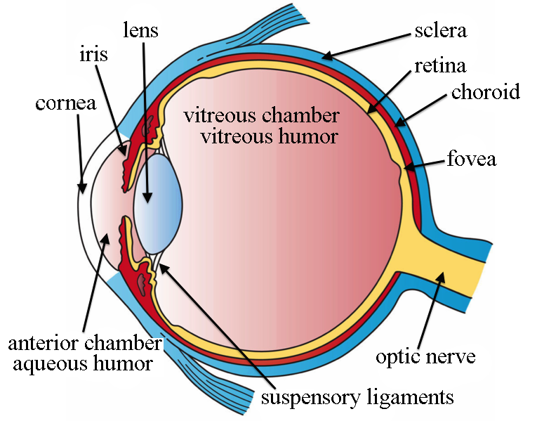
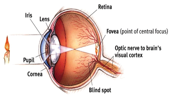
AP Psychology Anatomy of the Eye Diagram | Quizlet AP Psychology Diagram - Ear, Eye diagram: AP Psychology, Biological Psychology: Brain Diagram, Biological Psychology: Brain Diagram 69 Terms. lilmrslrobz TEACHER; Flickr Creative Commons Images. Some images used in this set are licensed under the Creative Commons through Flickr.com. AP Psych Unit 3 Notes: Visual Anatomy Review | Fiveable First, light passes through the cornea, a thin tissue that protects the eye and bends light to provide focus. 2. Next, light passes through the pupil, a small opening controlled by the iris. The iris is a colored muscle that constricts (gets smaller) or dilates (gets larger) based on light intensity.
PDF Ap Psychology Chapter 4: Sensation [AP PSYCHOLOGY CHAPTER 4: SENSATION] The Ear & Auditory Sensation Using textbook pg 111-118, identify the terms and concepts & answer the questions associated with the auditory sensations. • Sound: A repeated fluctuation, a rising and falling, in the pressure of air, water, or some other substance called a medium.

Eye diagram ap psychology
AP Psychology Eye diagram Flashcards - Quizlet AP Psychology Eye diagram STUDY Flashcards Learn Write Spell Test PLAY Match Gravity Retina Click card to see definition 👆 The light-sensitive inner surface of the eye, containing the receptor rods and cones plus layers of neurons that begin the processing of visual information. Click again to see term 👆 1/8 Previous ← Next → Flip Space Psychology- Eye & Ear Diagram Flashcards | Quizlet Eye Diagram Iris Muscles controls the size of the pupil. Pupil Iris opening that changes size depending on the amount of light in the environment. Cornea Bends light waves so the image can be focused on the retina. Lens Changes shape to bring objects into focus. Retina Contains photoreceptor cells. Fovea AP Psychology chapter 4: Sensation Label and shade the diagram of the eye below (only label and shade the bolded parts). Describe the functions of each eye part in the chart that follows. Eye ...4 pages
Eye diagram ap psychology. Structure and Functions of Human Eye with labelled Diagram Human Eye Diagram: Contrary to popular belief, the eyes are not perfectly spherical; instead, it is made up of two separate segments fused together. Explore: Facts About The Eye To understand more in detail about our eye and how our eye functions, we need to look into the structure of the human eye. PDF Mnemonic Devices for the Eye and Ear By Michael A. Britt ... along the back of the eye and it contains the rods, cones, bipolar and ganglion cells. Use "red tin" as your mnemonic and imagine that the back of your eye is covered with red tin. Fovea: is a spot in the eye that is directly behind the lens. There is a very high concentration of cones in this area which means that images t Quia You will diagram out the following 1. Diagram out the major parts of the eye with a brief description of their function 2. Diagram out an example of Nearsightedness, Farsightedness, and Perfect 20/20 Vision 3. Diagram out a close up of the Photoreceptors with explanation on how these w o rk. 4. Diagram out the steps in the process of Transduction. 2) Process of Transduction - Hook AP Psychology 4B 2 When light first enters the eye, it passes through the cornea and the pupil and hits the lens. This lens is converging and helps to focus the incoming object. An object is focused by the convex...
AP Psychology Ch.1-5 Cheat Sheet - Cheatography.com AP Psychology Ch.1-5 Cheat Sheet by MelissaM021004 via cheatography.com/122490/cs/22762/ People Albert Bandura Added a cognitive slant to behavi orism by resear ching violence and aggression B.F. Skinner Believed that internal mental events could only be studied scient ifi cally or not at all; Skinner box Carl Rogers Developed person -ce ntered Vision | Introduction to Psychology - Lumen Learning The anatomy of the eye is illustrated in this diagram. After passing through the pupil, light crosses the lens, a curved, transparent structure that serves to provide additional focus. The lens is attached to muscles that can change its shape to aid in focusing light that is reflected from near or far objects. Vision - AP Psychology Community There are essentially four steps to vision. First we have to gather light into our eye. The light has to be channeled to the back of the eye. Transduction occurs. The information goes to our brain where we interpret it. Let's break down the 4 steps in a little more detail. Step One: Gathering Light Unit 4 - AP PSYCHOLOGY Unit 4 ppt. File Size: 7020 kb. File Type: pdf. Download File. Optional Study Guide - hehe for the kids who actually look at the class website. File Size: 14 kb.
Brain: Ultimate Guide to the Brain for AP® Psychology For the AP® Psychology test, it is most important that you understand the functions I just listed and be familiar with the reticular formation. This structure controls our body's general arousal and our ability to focus, and it is a collection of cells spread throughout the midbrain. The Hindbrain Myers, Psychology for AP, 2e | Student Resources Module 9 Flip It Video - Action Potential. Module 10 Flip It Video - The Reflex Arc. Module 10 Flip It Video - Structure of the Nervous System. Module 11 Flip It Video - Limbic System. Module 12 Flip It Video - Structure and Function of the Cortex. Module 13 Flip It Video - Split-Brain Research. Label the Brain Worksheets (SB11585) - SparkleBox | Human ... Sep 30, 2018 - A set of diagrams of the brain. Includes a labelled versions for class discussion, as well as worksheets for pupils to label themselves (colour and black and white). AP Psychology 19-20 - Ms. Solomon All work assigned on Monday at 8am and due by Thursday at 4pm 1- Complete the Unit 1 Progress Check on AP Classroom 2- Complete the Unit 2 Progress Check on AP Classroom 3- Complete three viewing guides for review videos on YouTube by College Board (see TEAMs for directions and viewing guide) Virtual Learning Week #2 Assignments
AP Psych Eye Diagram Diagram - Quizlet AP Psych Eye Diagram STUDY Learn Flashcards Write Spell Test PLAY Match Gravity Created by Josiah728 Terms in this set (9) Cornea Clear, curved bulge of tissue on the front of the eye that bends light rays to begin focusing them and protects the eye (lots of nerve endings) Iris
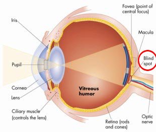
PDF Eye Anatomy Handout - National Eye Institute of light entering the eye. Lens: The lens is a clear part of the eye behind the iris that helps to focus light, or an image, on the retina. Macula: The macula is the small, sensitive area of the retina that gives central vision. It is located in the center of the retina. Optic nerve: The optic nerve is the largest sensory nerve of the eye.
Unit IV: Sensation and Perception - AP Psychology Eye Diagram Assignment. CHAPTER OBJECTIVES. After completing their study of this chapter, students should be able to: Contrast sensation and perception, and explain the difference between bottom-up and top-down processing. Distinguish between absolute and difference thresholds, and discuss whether we can sense and be affected by subliminal ...
AP_Eye_Diagram - Name: _ Parts of the Eye and their ... View Notes - AP_Eye_Diagram from AP PSYCH AP Psych at Deep Run High. Name: _ Parts of the Eye and their Functions Parts of the Eye 1. Cornea: _ _ _ _ _ 2. Pupil: _ _ _ _ _ 3. Iris: _ _ _ _ _ 4. Lens:
Human Eye Diagram Teaching Resources - Teachers Pay Teachers The Human Eye Overview Reading Comprehension and Diagram Worksheet. by. Teaching to the Middle. 58. $1.50. Zip. This passage briefly describes the human eye (900-1000 Lexile). 14 questions (matching and multiple choice) assess students' understanding. Students label a diagram of 6 parts of the eye.
AP Psychology -- Parts of the Eye Flashcards | Quizlet Clear, smooth, part that protects the eyeball and bends the might into the pupil. Lens The transparent structure behind the pupil that changes cap to help focus images on the retina. Pupil Center of the eye, allows light to enter (Controls amount of light is allowed into the eye) Iris Muscle that surrounds the pupil (The colored part). Blind spot?
PDF Parts of the Eye - National Eye Institute Eye Diagram Handout Author: National Eye Health Education Program of the National Eye Institute, National Institutes of Health Subject: Handout illustrating parts of the eye Keywords: parts of the eye, eye diagram, vitreous gel, iris, cornea, pupil, lens, optic nerve, macula, retina Created Date: 12/16/2011 12:39:09 PM
PDF Ultimate Study Guide for AP Psychology Ultimate Study Guide for AP Psychology ... • how light waves travel through eye and get transduced • major eye anatomy • (including rods and cones - see diagrams ) • feature detectors in occipital lobe in brain • why we have a blind spot • trichromatic color theory • opponent-process color theory ...
Eye_and_Ear_Anatomy_Quiz.pdf - AP Psychology Name Learning ... AP Psychology Name: _____ Part III The Eye (13-16) Directions: Complete the following fill in the blank questions regarding the specialized retinal cells responsible for visual transduction by selecting the best choice. Cones are specialized cells located primarily in the middle of the retina called the __fovea____.
Copy_of_Eye_and_Ear_Anatomy_Quiz.docx - AP Psychology Name ... AP Psychology Name: _____ Eye and Ear Anatomy Learning Target: Describe the sensory process for vision and audition including the specific nature of energy transduction, relevant anatomical structures, and the specialized pathway to the brain.
Human Eye Anatomy Quiz - Sporcle Top Quizzes with Similar Tags. Human Bones 90. Human Muscle Anatomy 67. Head-to-Toe Blitz 44. Human Heart Anatomy 25. Human Bones by Any Three Letters 24. Pop Quiz: Anatomy 12. Human Cranial Vault 8. Eye or Die 3.
AP Psychology chapter 4: Sensation Label and shade the diagram of the eye below (only label and shade the bolded parts). Describe the functions of each eye part in the chart that follows. Eye ...4 pages
Psychology- Eye & Ear Diagram Flashcards | Quizlet Eye Diagram Iris Muscles controls the size of the pupil. Pupil Iris opening that changes size depending on the amount of light in the environment. Cornea Bends light waves so the image can be focused on the retina. Lens Changes shape to bring objects into focus. Retina Contains photoreceptor cells. Fovea
AP Psychology Eye diagram Flashcards - Quizlet AP Psychology Eye diagram STUDY Flashcards Learn Write Spell Test PLAY Match Gravity Retina Click card to see definition 👆 The light-sensitive inner surface of the eye, containing the receptor rods and cones plus layers of neurons that begin the processing of visual information. Click again to see term 👆 1/8 Previous ← Next → Flip Space


Komentar
Posting Komentar